. 2012 Mar;347(3):701-11.
doi: 10.1007/s00441-011-1215-5. Epub 2011 Aug 11.

Scaffold-free culture of mesenchymal stem cell spheroids in suspension preserves multilineage potential

Affiliations

Scaffold-free culture of mesenchymal stem cell spheroids in suspension preserves multilineage potential

Priya R Baraniak et al. Cell Tissue Res. 2012 Mar.

Abstract

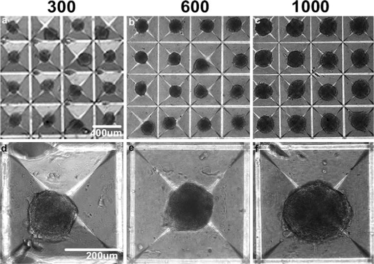
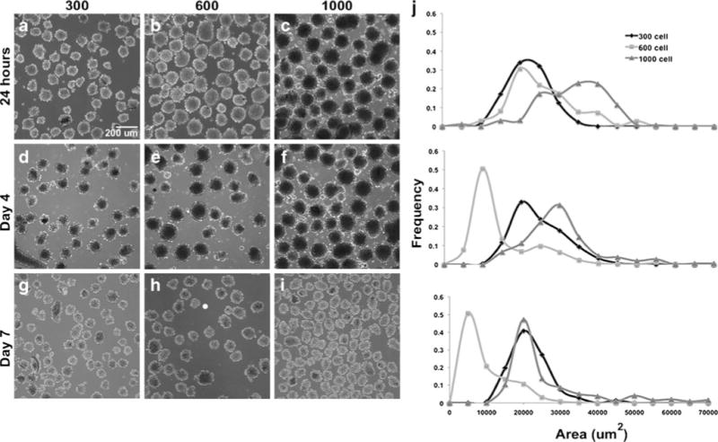
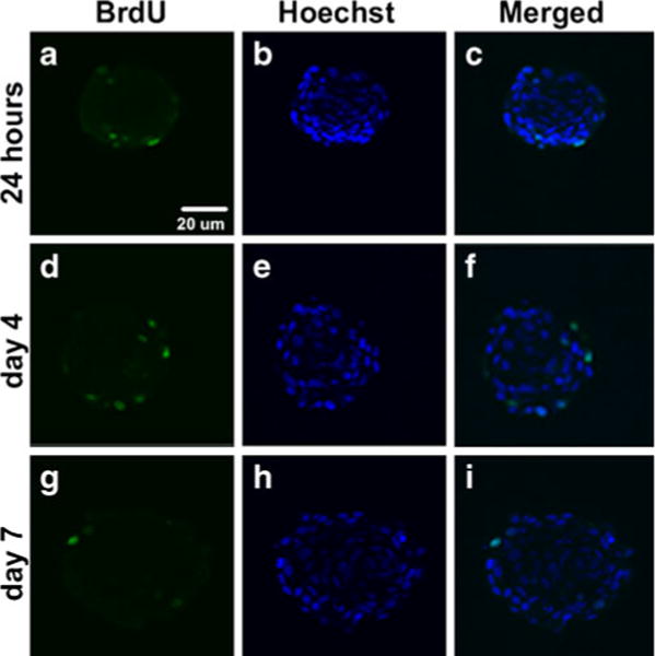
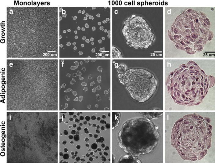
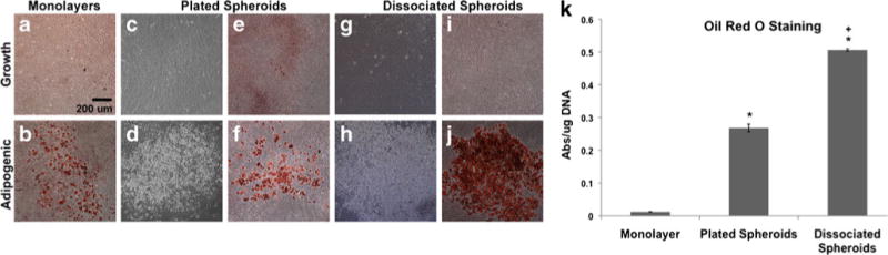
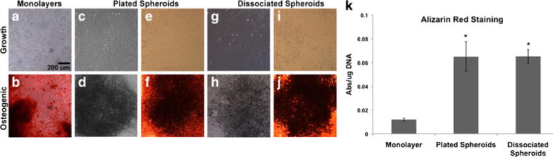
While traditional cell culture methods have relied on growing cells as monolayers, three-dimensional (3D) culture systems can provide a convenient in vitro model for the study of complex cell-cell and cell-matrix interactions in the absence of exogenous substrates and may benefit the development of regenerative medicine strategies. In this study, mesenchymal stem cell (MSC) spheroids, or "mesenspheres", of different sizes, were formed using a forced aggregation technique and maintained in suspension culture for extended periods of time thereafter. Cell proliferation and differentiation potential within mesenspheres and dissociated cells retrieved from spheroids were compared to conventional adherent monolayer cultures. Mesenspheres maintained in growth medium exhibited no evidence of cell necrosis or differentiation, while mesenspheres in differentiation media exhibited differentiation similar to conventional 2D culture methods based on histological markers of osteogenic and adipogenic commitment. Furthermore, when plated onto tissue culture plates, cells that had been cultured within mesenspheres in growth medium recovered morphology typical of cells cultured continuously in adherent monolayers and retained their capacity for multi-lineage differentiation potential. In fact, more robust matrix mineralization and lipid vacuole content were evident in recovered MSCs when compared to monolayers, suggesting enhanced differentiation by cells cultured as 3D spheroids. Thus, this study demonstrates the development of a 3D culture system for mesenchymal stem cells that may circumvent limitations associated with conventional monolayer cultures and enhance the differentiation potential of multipotent cells.


Figures

Fig. 1
Fig. 1
Mesensphere formation. Mesenspheres of controlled size, incorporating a 300, b 600, or c 1,000 cells each, were formed using forced-aggregation followed by MSC overnight incubation in Aggrewell™ inserts. df The relative size of the spheroids formed overnight correlated to the average number of cells entrapped within the individual wells. Scale bars (ac) 400 μm, (df) 200 μm
Fig. 2
Fig. 2
Mesensphere maintenance and size analysis. Following overnight mesensphere formation, spheroids were transferred to suspension culture on a rotary orbital shaker for subsequent analyses. ai Uniform starting populations of spheroids were obtained at each of the cell densities examined and homogeneous populations were maintained for extended periods of time (up to 7 days shown) in suspension without significant agglomeration. j Morphometric size analyses confirmed that homogeneous mesensphere populations could be maintained through at least 7 days of suspension culture. The 300-cell spheroids initially increased in size while the 600- and 1,000-cell spheroid sizes decreased slightly. By day 7 of culture, the 1,000-cell spheroid average area was comparable to the 300-cell spheroid average area. Scale bar 200 μm
Fig. 3
Fig. 3
Cell proliferation within mesenspheres up to 7 days of culture. Mesenspheres in suspension culture were pulsed with BrdU to confirm MSC proliferation within spheroids. ai BrdU staining indicated the presence of cycling cells within spheroids through 7 days of suspension culture. a, d, g However, BrdU+ cells were observed mainly at the periphery of spheroids and comprised only a small percentage (<5%) of the total number of cells. Scale bar 20 μm
Fig. 4
Fig. 4
Maintenance of mesensphere differentiation potential in suspension culture. Monolayers and mesenspheres maintained under growth and differentiation conditions were examined using phase contrast microscopy for signs of differentiation. Mesenspheres were also histologically examined for differences in morphology. ac Monolayers and mesenspheres maintained in growth medium (up to 16 days of culture) did not demonstrate lipid vacuole formation or matrix mineralization. eg Lipid vacuole formation was evident in adipogenic monolayers by day 28 of differentiation but could not be discerned in adipogenic mesenspheres at day 14. b, c, f, g Mesenspheres maintained in adipogenic medium resembled those in growth medium upon microscopic examination. ik Matrix mineralization was evident in both osteogenic monolayers and mesenspheres by day 14 of differentiation (opaque regions). d, h, l H&E staining of mesenspheres did not demonstrate any major differences in morphology between conditions. Scale bars (a, b, e, f, i, j) 200 μm, (c, d, g, h, k, l) 25 μm
Fig. 5
Fig. 5
Mesensphere histological examination. Following suspension culture, mesenspheres were histologically examined for ECM deposition and markers of adipogenic and osteogenic differentiation. a, d Collagen deposition, assayed using Masson’s trichrome staining, was not evident in growth or adipogenic spheroids. g However, Masson’s trichrome staining indicated the presence of collagen (stained blue, arrows) within the ECM of osteogenic mesenspheres at day 14 of differentiation. b, e, h Positive von Kossa staining for calcium mineral deposits (stained black, arrows) was evident within osteogenic mesenspheres but not others following 14 days of differentiation. f Lipid vacuole formation (stained red) within adipogenic mesenspheres was confirmed by Nile Red staining at day 14 of differentiation. c, i Some lipid vacuole formation was evident in growth mesenspheres but none was evident in osteogenic mesenspheres. Scale bar 20 μm
Fig. 6
Fig. 6
Adipogenic differentiation potential of MSCs following 3D culture. Following 7 days of suspension culture, MSCs were recovered from mesenspheres, plated onto TCPS and maintained under growth or adipogenic culture conditions. The adipogenic differentiation potential of recovered cells was compared to that of conventional monolayer cultures. Brightfield (a, b, e, f, i, j) and phase (c, d, g, h) images of Oil Red O-stained MSCs were obtained following differentiation. a, c, e, g, i Lipid vacuole formation was not evident in any cultures maintained under growth conditions. b, d, f, h, j Lipid vacuoles were more widespread within cultures recovered from mesenspheres following 14 days of differentiation than in monolayer cultures after 28 days of differentiation. Scale bar 200 μm. k Quantitative staining for adipogenesis exhibited significant differences between monolayer cultures and cells recovered from mesenspheres. Cells recovered from mesenspheres stained more robustly for Oil Red O than monolayers at day 14 of differentiation, with plated spheroids exhibiting a greater than 20-fold increase (*p< 0.0001) and dissociated spheroids exhibiting a greater than 40-fold increase (*p<0.0001) in the amount of lipid vacuole formation compared to monolayers. Dissociated mesenspheres also underwent greater adipogenesis (+p<0.0001) than plated spheroids
Fig. 7
Fig. 7
Osteogenic differentiation potential of MSCs following 3D culture. Following 7 days of suspension culture, MSCs were recovered from mesenspheres, plated onto TCPS and maintained under growth or osteogenic culture conditions. The osteogenic differentiation potential of recovered cells was compared to that of conventional monolayer cultures. Brightfield (a, b, e, f, i, j) and phase (c, d, g, h) images of Alizarin Red stained MSCs were obtained following differentiation. a, c, e, g, i Mineralization was not evident in any cultures maintained under growth conditions. b, d, f, h, j Mineralized regions were widely visible throughout all monolayer and recovered MSC osteogenic cultures by 14 days of differentiation. Scale bar 200 μm. k Quantitative staining for calcium deposits exhibited significant differences between monolayer cultures and cells recovered from mesenspheres. Cells recovered from mesenspheres stained more robustly for Alizarin Red than monolayers at day 14 of differentiation, with plated spheroids and dissociated cells exhibiting a greater than five-fold increase (*p<0.0001) in the amount of matrix mineralization compared to monolayers

References

    1. Dominici M, Le Blanc K, Mueller I, Slaper-Cortenbach I, Marini F, Krause D, Deans R, Keating A, Prockop D, Horwitz E. Minimal criteria for defining multipotent mesenchymal stromal cells. The International Society for Cellular Therapy position statement. Cytotherapy. 2006;8(4):315–317. - PubMed
    1. Pittenger MF. Mesenchymal stem cells from adult bone marrow. Methods Mol Biol. 2008;449:27–44. - PubMed
    1. Pittenger MF, Mackay AM, Beck SC, Jaiswal RK, Douglas R, Mosca JD, Moorman MA, Simonetti DW, Craig S, Marshak DR. Multilineage potential of adult human mesenchymal stem cells. Science. 1999;284(5411):143–147. - PubMed
    1. Baraniak PR, McDevitt TC. Stem cell paracrine actions and tissue regeneration. Regen Med. 2010;5(1):121–143. - PMC - PubMed
    1. Bonab MM, Alimoghaddam K, Talebian F, Ghaffari SH, Ghavamzadeh A, Nikbin B. Aging of mesenchymal stem cell in vitro. BMC Cell Biol. 2006;7:14. - PMC - PubMed

Publication types

LinkOut - more resources