

Kyrieleis plaques in cytomegalovirus retinitis
- PMID: 21833831
- PMCID: PMC3223340
- DOI: 10.1007/s12348-011-0033-y
Kyrieleis plaques in cytomegalovirus retinitis
Abstract
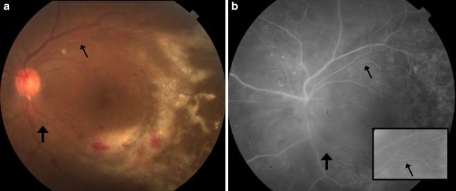
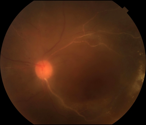
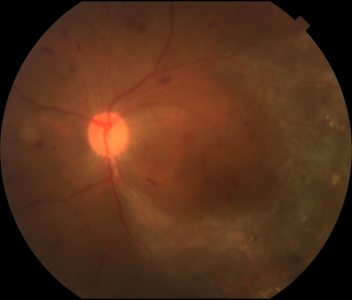
Purpose: The purpose of this study is to report a case of Kyrieleis plaques (segmental retinal periarteritis) associated with cytomegalovirus (CMV) retinitis.
Methods: A 47-year-old female with recently diagnosed human immunodeficiency virus and a CD4 count of 55 cells/µl presented with decreased vision and floaters in her left eye. Ophthalmic examination revealed an advancing border of white granular CMV retinitis extending into the macula. Intraocular aqueous specimen contained 420,000 copies/ml of CMV DNA by polymerase chain reaction. The patient was treated with intravitreal foscarnet and oral valganciclovir.
Results: Kyrieleis plaques involving the retinal arteries were noted on presentation and increased during the first 6 weeks of treatment as the retinitis faded. The plaques on fluorescein angiography did not leak fluorescein dye and slowly faded over 5 months.
Conclusions: Kyrieleis plaques can be seen in the setting of CMV retinitis. These plaques can be differentiated from vascular sheathing and frosted branch angiitis by its occurrence only in the retinal arteries and the absence of leakage of fluorescein dye.
Figures
References
-
- Kyrieleis W. Uber atypische gerfaesstuberkulose der netzhaut. Arch Augenheilkd. 1933;107:182–190.
-
- Griffin AO, Bodian M. Segmental retinal periarteritis; a report of three cases. Am J Ophthalmol. 1959;47:544–548. - PubMed
-
- Schwartz PL. Segmental retinal periarteritis as a complication of toxoplasmosis. Ann Ophthalmol. 1977;9:157–162. - PubMed
LinkOut - more resources
Full Text Sources
Research Materials
