Review
. 2011 Nov-Dec;3(6):620-46.
doi: 10.1002/wnan.158. Epub 2011 Aug 10.

Emerging applications of nanotechnology for the diagnosis and management of vulnerable atherosclerotic plaques

Affiliations
Review

Emerging applications of nanotechnology for the diagnosis and management of vulnerable atherosclerotic plaques

Shann S Yu et al. Wiley Interdiscip Rev Nanomed Nanobiotechnol. 2011 Nov-Dec.

Abstract

An estimated 16 million people in the United States have coronary artery disease (CAD), and approximately 325,000 people die annually from cardiac arrest. About two-thirds of unexpected cardiac deaths occur without prior recognition of cardiac disease. A vast majority of these deaths are attributable to the rupture of 'vulnerable atherosclerotic plaques'. Clinically, plaque vulnerability is typically assessed through imaging techniques, and ruptured plaques leading to acute myocardial infarction are treated through angioplasty or stenting. Despite significant advances, it is clear that current imaging methods are insufficiently capable for elucidating plaque composition--which is a key determinant of vulnerability. Further, the exciting improvement in the treatment of CAD afforded by stenting procedures has been buffered by significant undesirable host-implant effects, including restenosis and late thrombosis. Nanotechnology has led to some potential solutions to these problems by yielding constructs that interface with plaque cellular components at an unprecedented size scale. By leveraging the innate ability of macrophages to phagocytose nanoparticles, contrast agents can now be targeted to plaque inflammatory activity. Improvements in nano-patterning procedures have now led to increased ability to regenerate tissue isotropy directly on stents, enabling gradual regeneration of normal, physiologic vascular structures. Advancements in immunoassay technologies promise lower costs for biomarker measurements, and in the near future, may enable the addition of routine blood testing to the clinician's toolbox--decreasing the costs of atherosclerosis-related medical care. These are merely three examples among many stories of how nanotechnology continues to promise advances in the diagnosis and treatment of vulnerable atherosclerotic plaques.


Figures

FIGURE 1
FIGURE 1
Changes in mechanical properties of the vascular wall as a result of pathological vascular remodeling. (a) Ex vivo stress—strain curves of porcine plaque-laden versus healthy vascular tissue. All samples were taken from the left anterior descending coronary artery in juvenile pigs, with atherosclerosis induced via standard balloon angioplasty injury. Control samples were obtained from pigs without induced injury. (Reprinted with permission from Ref . Copyright 2003 ASME Publications.) (b) Schematic of changes in layer-by-layer stiffness during progression of pathological vascular remodeling. Pathological changes that decrease the lumen of a remodeling artery include intimal thickening and constrictive geometric remodeling of the wall, leading to a significant increase in the stiffness of inner layer, while vessel wall rupture that is resulted from artery expansion to increase the lumen for restoration of proper blood flow leads to decreased stiffness of middle and outer layers.
FIGURE 2
FIGURE 2
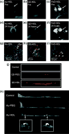
Multimodality imaging of atherosclerosis using nanocrystals encapsulated in high density lipoprotein. T1-weighted MR images of the aorta of apoE KO mice pre- (a and b) and 24 h postinjection (d and e) with Au-HDL or QD-HDL. Arrows indicate areas enhanced in the post images. (c and f) T2*-weighted images of an apoE KO mouse pre- and 24 h postinjection with FeO-HDL. (g–i) Confocal microscopy images of aortic sections of mice injected with nanocrystal HDL. Red is nanocrystal HDL, macrophages are green, and nuclei are blue. Yellow indicates colocalization of nanocrystal HDL with macrophages and is indicated by arrowheads. (j) Fluorescence image of aortas of mice injected with QD-HDL, QDPEG, and saline. (k) Ex vivo sagittal CT images of the aortas of mice injected with Au-HDL, Au-PEG, and saline. (Reprinted with permission from Ref . Copyright 2008 American Chemical Society)
FIGURE 3
FIGURE 3
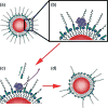
Dual-ligand, protease-activatable quantum dots for imaging applications. In ‘Proximity Activated Targeting’ (PAT) the targeting ligand (blue) is initially concealed (a) until proteolytic activity through tumor-secreted MMPs (purple) cleaves a peptide bridge (green) within long chain PEGs. Subsequent diffusion of the MMP and cleavage fragments (c) reveals the ligand-targeted construct only in the proximity of the tumor (d).
FIGURE 4
FIGURE 4
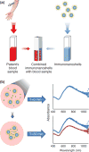
Nanoshell immunoassay. (a) In concept, the nanoshell immunoassay is designed to enable rapid and accurate analyte concentration measurements in whole blood, while minimizing dependency on long sample handling and instrumentation typified by standard immunoassays. (b) Absorbance spectra of nanoshells prior to versus 30 min after analyte addition. Peak absorbance is estimated at 725 nm. A decrease in material extinction coefficient is observed as a result of clustering of the nanoshells.
FIGURE 5
FIGURE 5
Bovine aortic endothelial cell adhesion and function on nanotextured versus conventional MP35N surfaces. Actin staining on a control dish (plastic), non-nanotextured and nanotextured MP35N surfaces. The presence of peri-junctional cortical bands of filamentous actin are clearly visible (arrows) on both the control and textured surfaces, but are not as pronounced on the nontextured surface, showing multiple layers of possibly aggregated cells. (Reprinted with permission from Ref . Copyright 2008 Elsevier)

Similar articles

Cited by

References

    1. Lloyd-Jones D, Adams RJ, Brown TM, Carnethon M, Dai S, De Simone G, Ferguson TB, Ford E, Furie K, Gillespie C, et al. Heart disease and stroke statistics—2010 update: a report from the American Heart Association. Circulation. 2010;121:e46–e215. - PubMed
    1. Insull W., Jr The pathology of atherosclerosis: plaque development and plaque responses to medical treatment. Am J Med. 2009;122:S3–S14. - PubMed
    1. Naghavi M, Falk E. Asymptomatic Atherosclerosis. New York, NY: Humana Press; 2010. From vulnerable plaque to vulnerable patient; pp. 13–38.
    1. Sousa JE, Serruys PW, Costa MA. New frontiers in cardiology: drug-eluting stents: part I. Circulation. 2003;107:2274–2279. - PubMed
    1. Finn AV, Nakazawa G, Joner M, Kolodgie FD, Mont EK, Gold HK, Virmani R. Vascular responses to drug eluting stents: importance of delayed healing. Arterioscler Thromb Vasc Biol. 2007;27:1500–1510. - PubMed

FURTHER READING

    1. Buxton DB. Current status of nanotechnology approaches for cardiovascular disease: a personal perspective. WIREs Nanomed Nanobiotechnol. 2009;1:149–155. - PubMed
    1. Jarzyna PA, Gianella A, Skajaa T, Knudsen G, Deddens LH, Cormode DP, Fayad ZA, Mulder WJM. Multifunctional imaging nanoprobes. WIREs Nanomed Nanobiotechnol. 2010;2:138–150. - PMC - PubMed
    1. Puskas JE, Muñoz-Robledo LG, Hoerr RA, Foley J, Schmidt SP, Evancho-Chapman M, Dong J, Frethem C, Haugstad G. Drug eluting stent coatings. WIREs Nanomed Nanobiotechnol. 2009;1:451–462. - PubMed

Publication types

MeSH terms

LinkOut - more resources