Review
. 2011 Nov 15;15(10):2699-710.
doi: 10.1089/ars.2011.4068. Epub 2011 Aug 11.

Organellar dysfunction in the pathogenesis of pancreatitis

Affiliations
Review

Organellar dysfunction in the pathogenesis of pancreatitis

Ilya Gukovsky et al. Antioxid Redox Signal. .

Abstract

Significance: Acute pancreatitis is an inflammatory disease of exocrine pancreas that carries considerable morbidity and mortality; its pathophysiology remains poorly understood. During the past decade, new insights have been gained into signaling pathways and molecules that mediate the inflammatory response of pancreatitis and death of acinar cells (the main exocrine pancreas cell type). By contrast, much less is known about the acinar cell organellar damage in pancreatitis and how it contributes to the disease pathogenesis.

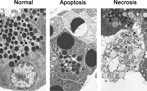
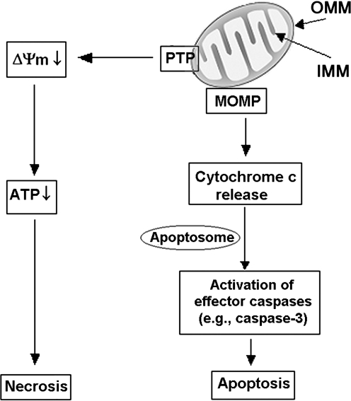
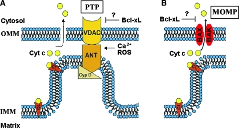
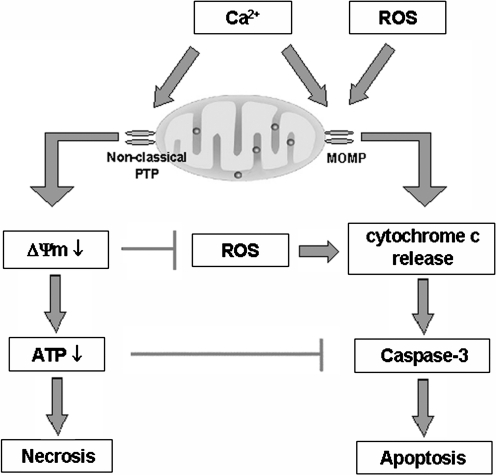
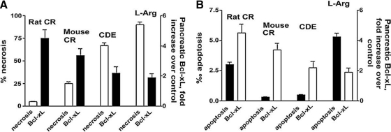
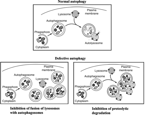
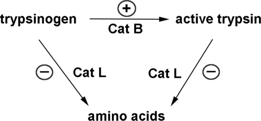
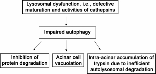
Recent advances: This review summarizes recent findings from our group, obtained on experimental in vivo and ex vivo models, which reveal disordering of key cellular organelles, namely, mitochondria, autophagosomes, and lysosomes, in pancreatitis. Our results indicate a critical role for mitochondrial permeabilization in determining the balance between apoptosis and necrosis in pancreatitis, and thus the disease severity. We further investigate how the mitochondrial dysfunction (and hence acinar cell death) is regulated by Ca(2+), reactive oxygen species, and Bcl-xL, in relation to specific properties of pancreatic mitochondria. Our results also reveal that autophagy, the principal cellular degradative, lysosome-driven pathway, is impaired in pancreatitis due to inefficient lysosomal function, and that impaired autophagy mediates two key pathological responses of pancreatitis-accumulation of vacuoles in acinar cells and the abnormal, intra-acinar activation of digestive enzymes such as trypsinogen.

Critical issues and future directions: The findings discussed in this review indicate critical roles for mitochondrial and autophagic/lysosomal dysfunctions in the pathogenesis of pancreatitis and delineate directions for detailed investigations into the molecular events that underlie acinar cell organellar damage.


Figures

FIG. 1.
FIG. 1.
Acinar cell apoptosis and necrosis in pancreatitis. Electron micrographs of normal rat pancreas and apoptotic and necrotic acinar cells in pancreatic tissue from rat with cerulein-induced pancreatitis.
FIG. 2.
FIG. 2.
Roles of mitochondria in apoptosis and necrosis. A critical event in cell death is mitochondrial membrane permeabilization, mediated by two permeability systems, the PTP and the mitochondrial outer MOMP (see text). PTP opening results in loss of the mitochondrial membrane potential (ΔΨm) and, ultimately, ATP depletion and necrosis. MOMP mediates the release into the cytosol of cytochrome c, triggering caspase activation cascade and subsequent apoptotic events. PTP, permeability transition pore; IMM, inner mitochondrial membrane; MOMP, membrane permeability system; OMM, outer mitochondrial membrane.
FIG. 3.
FIG. 3.
Mitochondrial permeability systems. (A) The PTP is a large nonselective channel the components of which include Cyp D, VDAC, and ANT. PTP opening is stimulated by Ca 2+ and ROS. (B) The MOMP channel is formed by the proapoptotic Bcl-2 proteins Bax and Bak. There is evidence that the prosurvival Bcl-xL protein inhibits both PTP and MOMP. Cyp D, cyclophilin D; VDAC, voltage-dependent anion channel; ANT, adenine nucleotide translocase; ROS, reactive oxygen species; Cyt c, cytochrome c; CL, cardiolipin. (To see this illustration in color the reader is referred to the web version of this article at www.liebertonline.com/ars).
FIG. 4.
FIG. 4.
The roles of Ca2+ and ROS in mitochondrial pathways of apoptosis and necrosis in pancreatitis. Ca2+ stimulates PTP opening resulting in ΔΨm loss, ATP depletion, and necrosis. ROS promote cytochrome c release through MOMP, resulting in caspase activation and apoptosis. On the other hand, Ca2+ has a dual effect on apoptosis. Ca2+ by itself stimulates cytochrome c release; however, Ca2+-induced depolarization, due to nonclassical properties of pancreatic mitochondrial PTP (see text), inhibits ROS production and thus cytochrome c release. In addition, the ATP decrease inhibits caspase activation. Thus, mitochondrial depolarization not only mediates necrosis but also limits apoptosis in pancreatitis, providing an explanation for the inverse correlation between the extent of acinar cell necrosis and apoptosis observed in experimental models of pancreatitis.
FIG. 5.
FIG. 5.
Increased pancreatic levels of Bcl-xL protect against necrosis in pancreatitis. The extent of Bcl-xL upregulation inversely correlates with acinar cell necrosis (A), but not apoptosis (B), in models of acute pancreatitis (Ref. , with permission). Pancreatitis was induced in rats and mice by administration of cerulein (CR), L-arginine (L-arg), or choline-deficient, ethionine-supplemented diet (CDE).
FIG. 6.
FIG. 6.
Schematic illustrating normal and defective macroautophagy, a sequential, lysosome-driven, adaptive process by which cells degrade cytoplasmic organelles and long-lived proteins (see text). This process begins when an isolation membrane (phagophore) wraps around material to be sequestered, for example, mitochondria, forming a double-membrane vacuole, the autophagosome. Autophagy progression/resolution (flux) proceeds through fusion of autophagosomes with lysosomes, generating autolysosomes, in which sequestered material is degraded. Two major mechanisms of defective autophagy are inhibition of the fusion between autophagosomes and lysosomes, resulting in accumulation of autophagosomes, and inefficient lysosomal degradation, resulting in accumulation of autolysosomes with partially degraded cargo.
FIG. 7.
FIG. 7.
Schematic illustrating the hypothesis (Ref. 51) that the pathological, intra-acinar trypsin accumulation results from disturbed equilibrium between the activities of cathepsin (Cat) B, which converts trypsinogen to trypsin, and CatL, which degrades both trypsin and trypsinogen. Such imbalance is caused by defective processing/maturation and activities of cathepsins in pancreatitis. The stimulatory and inhibitory effects of pancreatitis on CatB and CatL are shown by, respectively, (+) and (−) symbols.
FIG. 8.
FIG. 8.
Lysosomal/autophagic dysfunction mediates key pathological responses of pancreatitis.

References

    1. Adams JM. Cory S. Apoptosomes: engines for caspase activation. Curr Opin Cell Biol. 2002;14:715–720. - PubMed
    1. Adler G. Rohr G. Kern HF. Alteration of membrane fusion as a cause of acute pancreatitis in the rat. Dig Dis Sci. 1982;27:993–1002. - PubMed
    1. Aho HJ. Nevalainen TJ. Havia VT. Heinonen RJ. Aho AJ. Human acute pancreatitis: a light and electron microscopic study. Acta Pathol Microbiol Immunol Scand A. 1982;90:367–373. - PubMed
    1. Andreyev AY. Kushnareva YE. Starkov AA. Mitochondrial metabolism of reactive oxygen species. Biochemistry (Moscow) 2005;70:200–214. - PubMed
    1. Armstrong JS. Mitochondrial membrane permeabilization: the sine qua non for cell death. Bioessays. 2006;28:253–260. - PubMed

Publication types