. 2011 Aug 11;13(4):R79.
doi: 10.1186/bcr2928.

Transcriptome analysis of embryonic mammary cells reveals insights into mammary lineage establishment

Affiliations

Transcriptome analysis of embryonic mammary cells reveals insights into mammary lineage establishment

Olivia Wansbury et al. Breast Cancer Res. .

Abstract

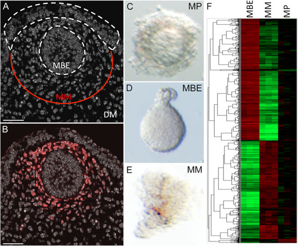
Introduction: The mammary primordium forms during embryogenesis as a result of inductive interactions between its constitutive tissues, the mesenchyme and epithelium, and represents the earliest evidence of commitment to the mammary lineage. Previous studies of embryonic mouse mammary epithelium indicated that, by mid-gestation, these cells are determined to a mammary cell fate and that a stem cell population has been delimited. Mammary mesenchyme can induce mammary development from simple epithelium even across species and classes, and can partially restore features of differentiated tissue to mouse mammary tumours in co-culture experiments. Despite these exciting properties, the molecular identity of embryonic mammary cells remains to be fully characterised.

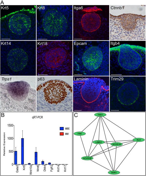
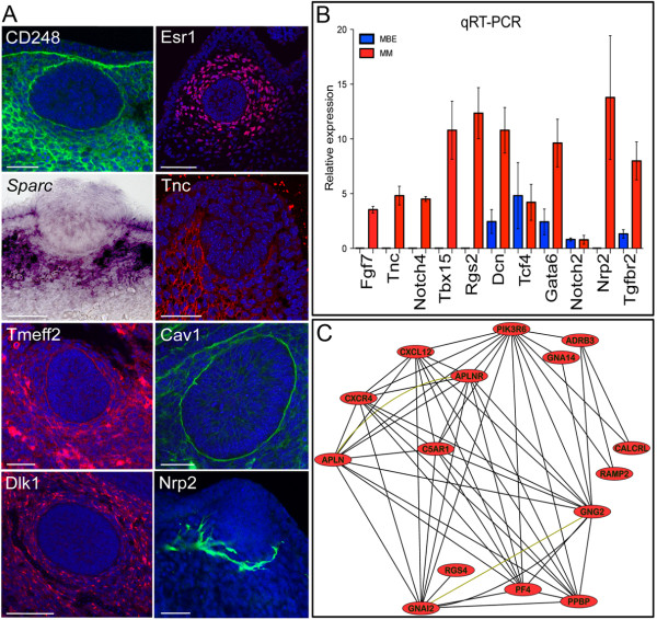
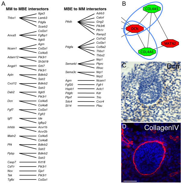
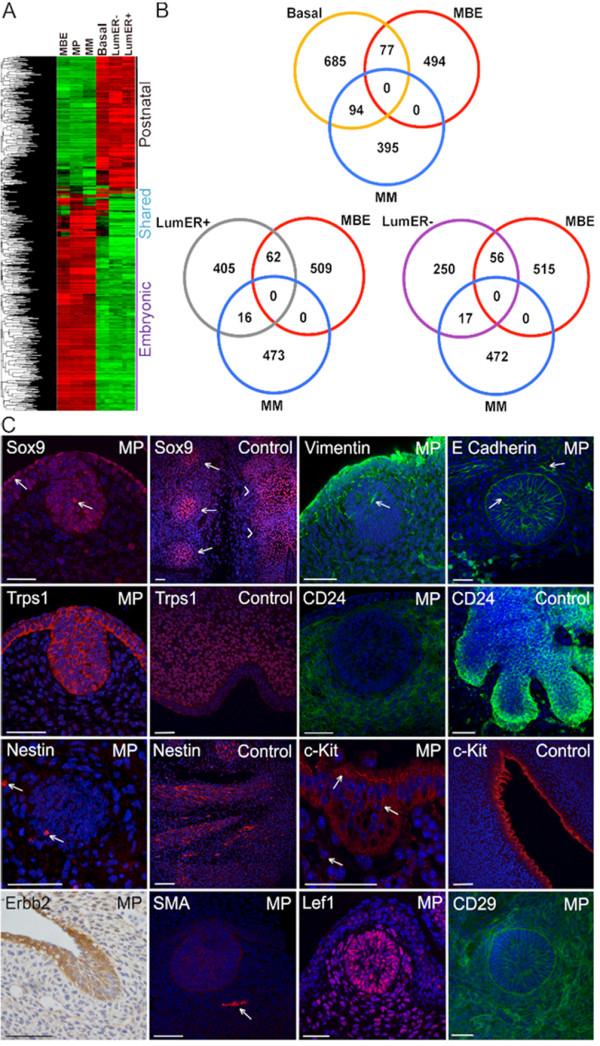
Methods: Here, we define the transcriptome of the mammary primordium and the two distinct cellular compartments that comprise it, the mammary primordial bud epithelium and mammary mesenchyme. Pathway and network analysis was performed and comparisons of embryonic mammary gene expression profiles to those of both postnatal mouse and human mammary epithelial cell sub-populations and stroma were made.

Results: Several of the genes we have detected in our embryonic mammary cell signatures were previously shown to regulate mammary cell fate and development, but we also identified a large number of novel candidates. Additionally, we determined genes that were expressed by both embryonic and postnatal mammary cells, which represent candidate regulators of mammary cell fate, differentiation and progenitor cell function that could signal from mammary lineage inception during embryogenesis through postnatal development. Comparison of embryonic mammary cell signatures with those of human breast cells identified potential regulators of mammary progenitor cell functions conserved across species.

Conclusions: These results provide new insights into genetic regulatory mechanisms of mammary development, particularly identification of novel potential regulators of mammary fate and mesenchymal-epithelial cross-talk. Since cancers may represent diseases of mesenchymal-epithelial communications, we anticipate these results will provide foundations for further studies into the fundamental links between developmental, stem cell and breast cancer biology.


Figures

Figure 1
Figure 1
Cell populations profiled to define the mammary primordial transcriptome. (A) E12.5 mammary primordium stained with DAPI; the amount of mammary mesenchyme (MM) and dermal mesenchyme (DM) left associated with the mammary bud epithelium (MBE) upon microdissection are indicated. (B) Immunofluorescence with ERα shows stains throughout the mesenchymal tissue that was isolated. Scale bar is 50 μm. (C) Microdissected mammary primordium (MP). (D) Mammary bud epithelium after tissue separation. (E) Mammary primordial mesenchyme after tissue separation. (F) Heatmap and supervised clustering of differential gene expression across mammary primordial tissues with probe sets with the most tissue-specific expression. Each horizontal line represents a probe set with red indicating high and green indicating low expression.
Figure 2
Figure 2
Validation and network analysis of mammary primordial epithelium microarray data. (A) Krt5, Krt14, Krt8, Krt18, Itga6/CD49f, and Epcam are expressed by many mammary primordial epithelial cells at high levels while some cells do not stain or express lower levels of these markers. All mammary primordial epithelial cells express p63. Laminin stains along the epidermal-mesenchymal boundary. Ctnnb1/β-catenin and Itgb4 are expressed at varying levels by cells within the primordia. Trim29 is expressed predominantly by basal cells. Scale bar is 50 μm. (B) qRT-PCR analysis of RNA from enzymatically separated E12.5 mammary bud epithelium (MBE) and mammary mesenchyme (MM). Gene RNA levels are expressed relative to Actb levels. Error bars ± s.e.m. Validation of eight mammary bud epithelium (MBE) specific genes showing enriched expression when compared to the mammary mesenchyme (MM). (C) A kinase-activin receptor sub-module is detected by network analysis. This sub-module forms part of the core network module generated using human orthologues of physically interacting genes characteristic of E12.5 mammary primordial epithelium. Black lines represent protein-protein interactions.
Figure 3
Figure 3
Validation and network analysis of mammary mesenchyme microarray data. (A) ERα and TnC stain the mammary mesenchyme. Most mammary mesenchymal cells express Sparc, except for those directly adjacent to the bud epithelium. Dlk1 and CD248/Endosialin stain all mesenchymal cells. Cav1 and Tmeff2 are expressed at higher levels by distinct cells. Nrp2 appears to be expressed by neural cells. Scale bar is 50 μm. (B) qRT-PCR analysis of RNA from enzymatically separated mammary bud epithelium (MBE) and mammary mesenchyme (MM). Gene RNA levels are expressed relative to Actb levels. Error bars ± s.e.m. Validation of nine MM specific genes enriched expression when compared to the MBE. Tcf4 did not show population specific expression by this analysis. (C) A sub-module comprised of several highly interconnected nodes representing G protein coupled receptor signalling and cytokines forms part of the core network module of mammary primordial mesenchyme. This core network was generated using human orthologs of directly interacting genes characteristic of the mammary mesenchyme. Black lines represent protein-protein interactions. Nodes that have been shown to interact in complexes are connected together by yellow-green lines.
Figure 4
Figure 4
Prospective mediators of epithelial-mesenchymal interactions identified by network analysis. (A) List of likely effectors of inductive signalling across the mesenchymal-epithelial boundary. In addition to ligand-receptor pairs, inter-connections between known signalling cascade components that could transmit signals across the mammary mesenchyme (MM) and the mammary bud epithelium (MBE) and vice versa are shown. (B) Mesenchymal Decorin (Dcn) is poised to interact with both epithelial CollagenIVa5 and CollagenIVa6 (Col4a5 and Col4a6). Red represents mesenchymal nodes. Green represents epithelial nodes. (C, D) Decorin and CollagenIV expression patterns suggest that these interconnected nodes represent feasible biological interactions that could modulate signals both within the extracellular matrix and basement membrane and across the mesenchymal-epithelial boundary. Scale bar is 50 μm.
Figure 5
Figure 5
Transcriptome analysis reveals similarities and differences between embryonic and postnatal mammary cell populations. (A) Heatmap and unsupervised cluster analysis of differential gene expression across postnatal mammary epithelial cells (MECs) and mammary primordial populations. Each horizontal line represents a probe set with red indicating high expression and green indicating low expression in the various tissue compartments. (B) Venn diagrams showing the number of shared and unique genes expressed within mammary primordial compartments compared to the three postnatal mouse MEC subpopulations. (C) Arrows indicate cells expressing nuclear Sox9 within the surface and primordial epithelium. Control tissue shows strong nuclear staining in the developing ribs (arrows) and spinal cord (arrowheads). Strong Vimentin expression is observed in the periderm, surface epithelium and mammary mesenchymal cells. Arrow indicates a mammary primordial epithelial cell expressing Vimentin. Most suprabasal cells of the mammary primordial epithelium express E-cadherin; less intense expression is observed in the periderm and surface epithelium above the mammary bud and some suprabasal cells (arrows). Trps1 stain is non-nuclear within the mammary bud epithelium. Vibrissae mesenchyme shows nuclear stain while some non-nuclear staining is observed in the epithelium at E12.5 (Control). Diffuse CD24 is observed in E12.5 mammary mesenchyme. At E16.5, the mammary bud epithelium and some cells within the mammary mesenchyme express CD24 (Control). A few Nestin-expressing cells are observed within both mammary epithelial and mesenchymal primordial cells. Strong Nestin staining is observed in embryonic nerves at E12.5. Some c-Kit positive cells are observed within the mammary bud epithelium and mesenchyme. Positive control is roof of brain ventricle at E12.5. Erbb2 expression is observed in mammary bud epithelial and some mesenchymal cells at E12.5. SMA is observed in a blood vessel (arrow) adjacent to the mammary bud epithelium, which does not express SMA. Both the mammary bud epithelial and mammary mesenchymal cells express Lef1 and CD29. Scale bar is 50 μm. MBE denotes mammary bud epithelium. MM denotes mammary mesenchyme. MP denotes mammary primordium. LumER- denotes luminal estrogen receptor negative. LumER+ denotes luminal estrogen receptor positive.

References

    1. Parmar H, Cunha GR. Epithelial-stromal interactions in the mouse and human mammary gland in vivo. Endocr Relat Cancer. 2004;11:437–458. doi: 10.1677/erc.1.00659. - DOI - PubMed
    1. Sakakura T, Nishizuka Y, Dawe CJ. Mesenchyme-dependent morphogenesis and epithelium-specific cytodifferentiation in mouse mammary gland. Science. 1976;194:1439–1441. doi: 10.1126/science.827022. - DOI - PubMed
    1. Klinowska TC, Streuli CH. Analyzing cell-ECM interactions in adult mammary gland by transplantation of embryonic mammary tissue from knockout mice. Methods Mol Biol. 2000;139:345–358. - PubMed
    1. Sakakura T, Nishizuka Y, Dawe CJ. Capacity of mammary fat pads of adult C3H/HeMs mice to interact morphogenetically with fetal mammary epithelium. J Natl Cancer Inst. 1979;63:733–736. - PubMed
    1. Kendrick H, Regan JL, Magnay FA, Grigoriadis A, Mitsopoulos C, Zvelebil M, Smalley MJ. Transcriptome analysis of mammary epithelial subpopulations identifies novel determinants of lineage commitment and cell fate. BMC Genomics. 2008;9:591. doi: 10.1186/1471-2164-9-591. - DOI - PMC - PubMed

Publication types

Substances