. 2011 Aug 11:3:19.
doi: 10.1186/2045-824X-3-19.

Expression of HES and HEY genes in infantile hemangiomas

Affiliations

Expression of HES and HEY genes in infantile hemangiomas

Omotinuwe Adepoju et al. Vasc Cell. .

Abstract

Background: Infantile hemangiomas (IHs) are the most common benign tumor of infancy, yet their pathogenesis is poorly understood. IHs are believed to originate from a progenitor cell, the hemangioma stem cell (HemSC). Recent studies by our group showed that NOTCH proteins and NOTCH ligands are expressed in hemangiomas, indicating Notch signaling may be active in IHs. We sought to investigate downstream activation of Notch signaling in hemangioma cells by evaluating the expression of the basic HLH family proteins, HES/HEY, in IHs.

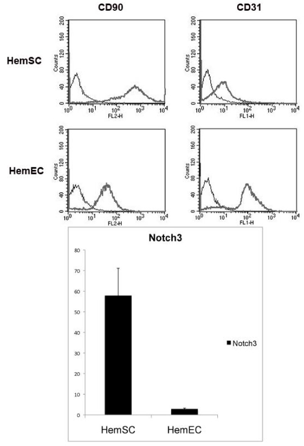
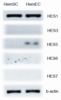
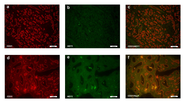
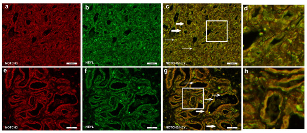
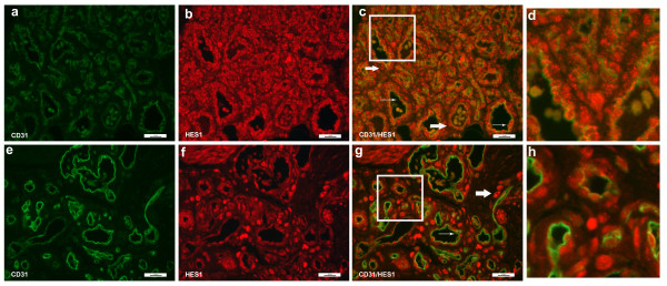
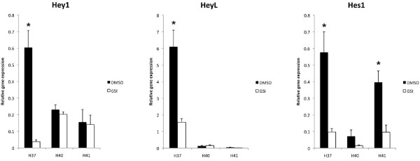
Materials and methods: HemSCs and hemangioma endothelial cells (HemECs) are isolated from freshly resected hemangioma specimens. Quantitative RT-PCR was performed to probe for relative gene transcript levels (normalized to beta-actin). Immunofluorescence was performed to evaluate protein expression. Co-localization studies were performed with CD31 (endothelial cells) and NOTCH3 (peri-vascular, non-endothelial cells). HemSCs were treated with the gamma secretase inhibitor (GSI) Compound E, and gene transcript levels were quantified with real-time PCR.

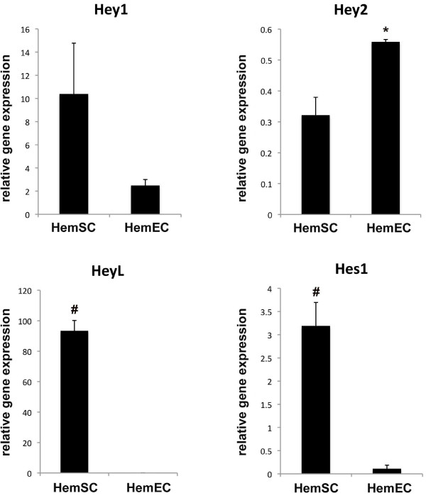
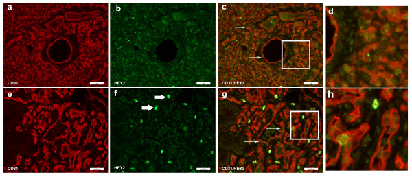
Results: HEY1, HEYL, and HES1 are highly expressed in HemSCs, while HEY2 is highly expressed in HemECs. Protein expression evaluation by immunofluorescence confirms that HEY2 is expressed by HemECs (CD31+ cells), while HEY1, HEYL, and HES1 are more widely expressed and mostly expressed by perivascular cells of hemangiomas. Inhibition of Notch signaling by addition of GSI resulted in down-regulation of HES/HEY genes.

Conclusions: HES/HEY genes are expressed in IHs in cell type specific patterns; HEY2 is expressed in HemECs and HEY1, HEYL, HES1 are expressed in HemSCs. This pattern suggests that HEY/HES genes act downstream of Notch receptors that function in distinct cell types of IHs. HES/HEY gene transcripts are decreased with the addition of a gamma-secretase inhibitor, Compound E, demonstrating that Notch signaling is active in infantile hemangioma cells.


Figures

Figure 1
Figure 1
FACS analysis. (a) FACS analysis results show that HemSCs are positive for CD90 and negative for CD31, consistent with previously published reports,[22] whereas HemECs are positive for CD31. NOTCH3 expression. (b) qPCR analysis showed that HemSCs express high levels of NOTCH3, while HemECs showed minimal to no NOTCH3 expression.
Figure 2
Figure 2
Survey PCR for HES gene. PCR analysis showed that HemSCs showed no expression of HES3, -5, -6, or 7; a very low level of expression was seen for HES5 in HemECs.
Figure 3
Figure 3
Quantitative PCR results of HES and HEY genes in HemSCs and HemECs. Quantitative PCR results showed that HES1, HEY1, and HEYL had higher transcript levels in HemSCs when compared to HemECs (*p<0.05, #p<0.01.) Gene transcript levels were expressed as relative to beta-actin levels. Results were done in triplicates at least in 2 separate experiments and representative of 2 different cell lines. Statistical analysis showed that the difference in transcript levels for HEY1 approached, but did not achieve, statistical significance (p = 0.08). However, the transcript levels were statistically different between HemSCs and HemECs: Hey2 (p = 0.02), HeyL (p = 0.002), and Hes1 (p = 0.008).
Figure 4
Figure 4
Immunofluorescence staining of CD31 and HEY1. Immunofluorescence staining CD31 (red) and HEY1 (green). There was no appreciable staining in the involuting hemangioma specimen (bottom panels, d-f). However, HEY1 was present in the proliferating hemangioma (a-c). Magnification, 40X.
Figure 5
Figure 5
Immunofluorescence staining of CD31 and HEY2. Immunofluorescence staining of CD31 (red) and HEY2 (green). Proliferating hemangioma, panels a-c; involuting hemangioma, panels e-g. HEY2 staining was seen in the endothelial cell nucleui, whereas CD31 staining was present in the cytoplasm. Therefore, while CD31 and HEY2 co-localized, the color did not overlap (thin arrows, panels c & g). Not all endothelial cells (red cytoplasmic staining) co-stained with HEY2, and there were non-endothelial cells that were HEY2 positive (Figure 5f, thick arrows, bright green). Magnification, 40X. Panels d, h: close up of panels 5c (proliferating hemangioma) and 5g (involuting hemangioma).
Figure 6
Figure 6
Immunofluorescence staining of NOTCH3 and HEYL. Immunofluorescence staining of NOTCH3 (green) and HEYL (red). Proliferating hemangioma, panels a-c; involuting hemangioma, panels e-g. NOTCH3+ cells are located in the perivascular regions and co-localize with HEYL (thick arrows, panels c & g). However, some endothelial cells also express HEYL (thin arrows, panels c & g). Magnification, 40×. Panels d, h: close up of Figure 6c (proliferating hemangioma) and 6g (involuting hemangioma). There were luminal cells that stained for HEYL (green, thick arrows) but not NOTCH3. The majority of HEYL positive cells co-localized with NOTCH3 (yellow).
Figure 7
Figure 7
Immunofluorescence staining of CD31 and HES1. Immunofluorescence staining of CD31 (green) and HES1 (red). Proliferating hemangioma, panels a-c; involuting hemangioma, panels e-g. HES1 was expressed in HemSCs and were localized in the peri-vascular cells (thick arrows, panels c & g). There were occasional endothelial cells that showed nuclear staining of HES1 and cytoplasmic staining of CD31 (thin arrows, panels d & f). Magnification, 40×. Panels d, h: close up of Figure 7c (proliferating hemangioma) and 7g (involuting hemangioma). There were occasional endothelial cells that expressed HES1 (red nuclei with green cytoplasm, thin arrows), while the majority of HES1+ cells (red nuclei) were located in non-endothelial perivascular cells.
Figure 8
Figure 8
Quantitative PCR results of HES/HEY genes after treatment with gamma secretase inhibitor. Transcript levels of HES/HEY genes in HemSCs are changed with administration of a gamma secretase inhibitor (GSI), Compound E. HEY1 (left) transcripts were universally decreased after GSI treatment, although only statistically significant for H37 (p = 0.01). HEYL (middle) transcripts were decreased in H37, (p = 0.02), but baseline transcript levels were too low for the other 2 cell lines for meaningful comparison. HES1 (right) transcripts were universally decreased and were statistically significant for H37 (p = 0.02) and H41 (p = 0.03). It was not significant for H40 (p = 0.1).

References

    1. Frieden IJ, Haggstrom AN, Drolet BA, Mancini AJ, Friedlander SF, Boon L, Chamlin SL, Baselga E, Garzon MC, Nopper AJ. et al.Infantile hemangiomas: current knowledge, future directions. Proceedings of a research workshop on infantile hemangiomas, April 7-9, 2005, Bethesda, Maryland, USA. Pediatr Dermatol. 2005;22(5):383–406. doi: 10.1111/j.1525-1470.2005.00102.x. - DOI - PubMed
    1. Mulliken JB, Fishman SJ, Burrows PE. Vascular anomalies. Curr Probl Surg. 2000;37(8):517–584. doi: 10.1016/S0011-3840(00)80013-1. - DOI - PubMed
    1. Artavanis-Tsakonas S, Rand MD, Lake RJ. Notch signaling: cell fate control and signal integration in development. Science. 1999;284(5415):770–776. doi: 10.1126/science.284.5415.770. - DOI - PubMed
    1. Alva JA, Iruela-Arispe ML. Notch signaling in vascular morphogenesis. Curr Opin Hematol. 2004;11(4):278–283. doi: 10.1097/01.moh.0000130309.44976.ad. - DOI - PubMed
    1. Iso T, Hamamori Y, Kedes L. Notch signaling in vascular development. Arterioscler Thromb Vasc Biol. 2003;23(4):543–553. doi: 10.1161/01.ATV.0000060892.81529.8F. - DOI - PubMed

LinkOut - more resources