

Comparison of long and short axis quantification of left ventricular volume parameters by cardiovascular magnetic resonance, with ex-vivo validation
- PMID: 21834992
- PMCID: PMC3169477
- DOI: 10.1186/1532-429X-13-40
Comparison of long and short axis quantification of left ventricular volume parameters by cardiovascular magnetic resonance, with ex-vivo validation
Abstract
Background: The purpose of the study was to compare the accuracy and evaluation time of quantifying left ventricular (LV), left atrial (LA) volume and LV mass using short axis (SAX) and long axis (LAX) methods when using cardiovascular magnetic resonance (CMR).
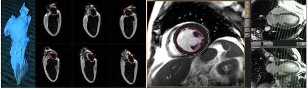
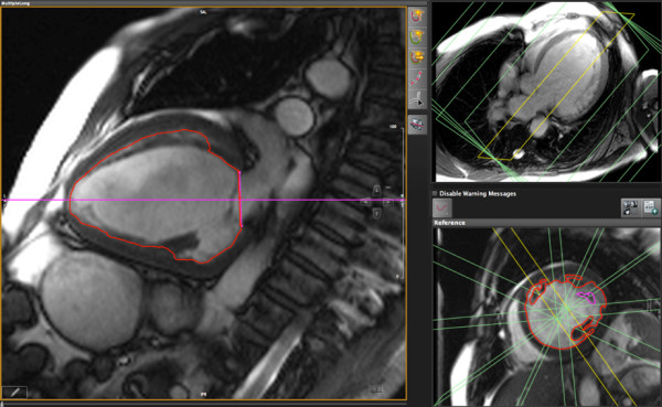
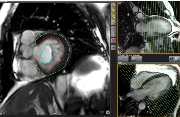
Materials and methods: We studied 12 explanted canine hearts and 46 patients referred for CMR (29 male, age 47 ± 18 years) in a clinical 1.5 T CMR system, using standard cine sequences. In standard short axis stacks of various slice thickness values in dogs and 8 mm slice thickness (gap 2 mm) in patients, we measured LV volumes using reference slices in a perpendicular, long axis orientation using certified software. Volumes and mass were also measured in six radial long axis (LAX) views.LV parameters were also assessed for intra- and inter-observer variability. In 24 patients, we also analyzed reproducibility and evaluation time of two very experienced (> 10 years of CMR reading) readers for SAX and LAX.
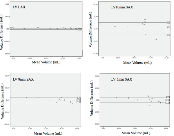
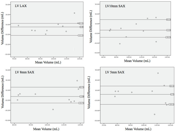
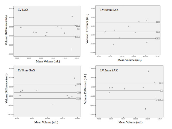
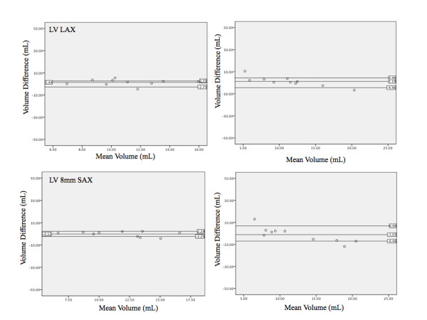
Results: In the explanted dog hearts, there was excellent agreement between ex vivo data and LV mass and volume data as measured by all methods for both, LAX (r² = 0.98) and SAX (r² = 0.88 to 0.98). LA volumes, however, were underestimated by 13% using the LAX views. In patients, there was a good correlation between all three assessed methods (r² ≥ 0.95 for all). In experienced clinical readers, left-ventricular volumes and ejection fraction as measured in LAX views showed a better inter-observer reproducibility and a 27% shorter evaluation time.
Conclusion: When compared to an ex vivo standard, both, short axis and long axis techniques are highly accurate for the quantification of left ventricular volumes and mass. In clinical settings, however, the long axis approach may be more reproducible and more time-efficient. Therefore, the rotational long axis approach is a viable alternative for the clinical assessment of cardiac volumes, function and mass.
Figures
Similar articles
-
Compressed sensing single-breath-hold CMR for fast quantification of LV function, volumes, and mass.JACC Cardiovasc Imaging. 2014 Sep;7(9):882-92. doi: 10.1016/j.jcmg.2014.04.016. Epub 2014 Aug 13. JACC Cardiovasc Imaging. 2014. PMID: 25129517
-
Transthoracic 3D Echocardiographic Left Heart Chamber Quantification Using an Automated Adaptive Analytics Algorithm.JACC Cardiovasc Imaging. 2016 Jul;9(7):769-782. doi: 10.1016/j.jcmg.2015.12.020. Epub 2016 Jun 15. JACC Cardiovasc Imaging. 2016. PMID: 27318718
-
Biplane versus short-axis measures of the left atrium and ventricle in patients with systolic dysfunction assessed by magnetic resonance.Clin Imaging. 2016 Sep-Oct;40(5):907-12. doi: 10.1016/j.clinimag.2016.04.015. Epub 2016 Apr 30. Clin Imaging. 2016. PMID: 27183139
-
Variation in left ventricular cardiac magnetic resonance normal reference ranges: systematic review and meta-analysis.Eur Heart J Cardiovasc Imaging. 2021 Apr 28;22(5):494-504. doi: 10.1093/ehjci/jeaa089. Eur Heart J Cardiovasc Imaging. 2021. PMID: 32460308 Free PMC article.
-
Reference ranges ("normal values") for cardiovascular magnetic resonance (CMR) in adults and children: 2020 update.J Cardiovasc Magn Reson. 2020 Dec 14;22(1):87. doi: 10.1186/s12968-020-00683-3. J Cardiovasc Magn Reson. 2020. PMID: 33308262 Free PMC article. Review.
Cited by
-
Clinical Performance and Role of Expert Supervision of Deep Learning for Cardiac Ventricular Volumetry: A Validation Study.Radiol Artif Intell. 2020 Jul 8;2(4):e190064. doi: 10.1148/ryai.2020190064. Radiol Artif Intell. 2020. PMID: 32797119 Free PMC article.
-
Three-dimensional Cardiomyocytes Structure Revealed By Diffusion Tensor Imaging and Its Validation Using a Tissue-Clearing Technique.Sci Rep. 2018 Apr 27;8(1):6640. doi: 10.1038/s41598-018-24622-6. Sci Rep. 2018. PMID: 29703900 Free PMC article.
-
Development and validation of AI-derived segmentation of four-chamber cine cardiac magnetic resonance.Eur Radiol Exp. 2024 Jul 12;8(1):77. doi: 10.1186/s41747-024-00477-7. Eur Radiol Exp. 2024. PMID: 38992116 Free PMC article.
-
Cardiotoxicity due to chemotherapy: role of cardiac imaging.Curr Cardiol Rep. 2015 Mar;17(3):564. doi: 10.1007/s11886-015-0564-1. Curr Cardiol Rep. 2015. PMID: 25648628 Review.
-
Comparative analysis of left ventricle function and deformation imaging in short and long axis plane in cardiac magnetic resonance imaging.Front Cardiovasc Med. 2024 May 2;11:1388171. doi: 10.3389/fcvm.2024.1388171. eCollection 2024. Front Cardiovasc Med. 2024. PMID: 38756751 Free PMC article.
References
-
- Hendel RC, Patel MR, Kramer CM, Poon M, Hendel RC, Carr JC, Gerstad NA, Gillam LD, Hodgson JM, Kim RJ, Kramer CM, Lesser JR, Martin ET, Messer JV, Redberg RF, Rubin GD, Rumsfeld JS, Taylor AJ, Weigold WG, Woodard PK, Brindis RG, Hendel RC, Douglas PS, Peterson ED, Wolk MJ, Allen JM, Patel MR. ACCF/ACR/SCCT/SCMR/ASNC/NASCI/SCAI/SIR 2006 appropriateness criteria for cardiac computed tomography and cardiac magnetic resonance imaging: a report of the American College of Cardiology Foundation Quality Strategic Directions Committee Appropriateness Criteria Working Group, American College of Radiology, Society of Cardiovascular Computed Tomography, Society for Cardiovascular Magnetic Resonance, American Society of Nuclear Cardiology, North American Society for Cardiac Imaging, Society for Cardiovascular Angiography and Interventions, and Society of Interventional Radiology. J Am Coll Cardiol. 2006;48:1475–97. doi: 10.1016/j.jacc.2006.07.003. - DOI - PubMed
-
- Hundley WG, Bluemke DA, Finn JP, Flamm SD, Fogel MA, Friedrich MG, Ho VB, Jerosch-Herold M, Kramer CM, Manning WJ, Patel M, Pohost GM, Stillman AE, White RD, Woodard PK. ACCF/ACR/AHA/NASCI/SCMR 2010 Expert Consensus Document on Cardiovascular Magnetic Resonance: A Report of the American College of Cardiology Foundation Task Force on Expert Consensus Documents. Circulation. 2010;121:2462–508. - PMC - PubMed
-
- Dulce MC, Mostbeck GH, Friese KK, Caputo GR, Higgins CB. Quantification of the left ventricular volumes and function with cine MR imaging: comparison of geometric models with three-dimensional data. Radiology. 1993;188:371–6. - PubMed
-
- Bloomer TN, Plein S, Radjenovic A, Higgins DM, Jones TR, Ridgway JP. Sivananthan MU Cine MRI using steady state free precession in the radial long axis orientation is a fast accurate method for obtaining volumetric data of the left ventricle. J Magn Reson Imaging. 2001;14:685–92. doi: 10.1002/jmri.10019. - DOI - PubMed
Publication types
MeSH terms
LinkOut - more resources
Full Text Sources
Miscellaneous
