

Functional connectivity and integrative properties of globus pallidus neurons
- PMID: 21835227
- PMCID: PMC3221766
- DOI: 10.1016/j.neuroscience.2011.07.050
Functional connectivity and integrative properties of globus pallidus neurons
Abstract
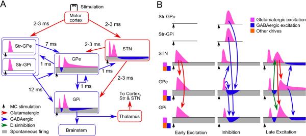
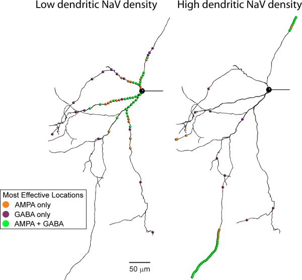
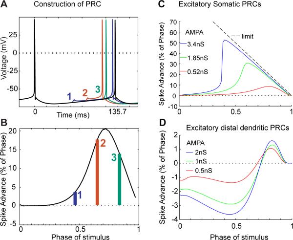
The globus pallidus consists of the external (GPe) and the internal (GPi) segments. The GPe and GPi have different functional roles. The GPe is located centrally within multiple basal ganglia feedforward and feedback connections. The GPi is an output nucleus of the basal ganglia. A complex interplay between intrinsic pacemaking conductances and the balance of glutamatergic and GABAergic input largely determines the rate and pattern of firing of pallidal neurons. The initial part of this article introduces recent findings made in vivo that are related to the roles of glutamatergic and GABAergic inputs in the control of pallidal activity. The latter part describes the roles of intrinsic mechanisms of GPe neurons in the integration of the synaptic inputs. The presence of dendritic voltage-gated sodium channels may allow the initiation of dendritic spikes, giving distal inputs on the long and thin GPe dendrites an opportunity to strongly shape spiking activity. Basal ganglia disorders including Parkinson's disease, hemiballismus, and dystonias are accompanied by increased irregularity and synchronized bursts of pallidal activity. These changes may be in part due to changes in the GABA release in the GPe and GPi, but also involve intrinsic cellular changes in pallidal neurons.
Copyright © 2011 IBRO. Published by Elsevier Ltd. All rights reserved.
Figures
Similar articles
-
Singing-related neural activity distinguishes two putative pallidal cell types in the songbird basal ganglia: comparison to the primate internal and external pallidal segments.J Neurosci. 2010 May 19;30(20):7088-98. doi: 10.1523/JNEUROSCI.0168-10.2010. J Neurosci. 2010. PMID: 20484651 Free PMC article.
-
Parvalbumin+ and Npas1+ Pallidal Neurons Have Distinct Circuit Topology and Function.J Neurosci. 2020 Oct 7;40(41):7855-7876. doi: 10.1523/JNEUROSCI.0361-20.2020. Epub 2020 Aug 31. J Neurosci. 2020. PMID: 32868462 Free PMC article.
-
Origins of GABA(A) and GABA(B) receptor-mediated responses of globus pallidus induced after stimulation of the putamen in the monkey.J Neurosci. 2006 Jun 14;26(24):6554-62. doi: 10.1523/JNEUROSCI.1543-06.2006. J Neurosci. 2006. PMID: 16775143 Free PMC article.
-
Active decorrelation in the basal ganglia.Neuroscience. 2013 Oct 10;250:467-82. doi: 10.1016/j.neuroscience.2013.07.032. Epub 2013 Jul 24. Neuroscience. 2013. PMID: 23892007 Free PMC article. Review.
-
Globus pallidus external segment.Prog Brain Res. 2007;160:111-33. doi: 10.1016/S0079-6123(06)60007-1. Prog Brain Res. 2007. PMID: 17499111 Review.
Cited by
-
Motor cortex can directly drive the globus pallidus neurons in a projection neuron type-dependent manner in the rat.Elife. 2019 Nov 12;8:e49511. doi: 10.7554/eLife.49511. Elife. 2019. PMID: 31711567 Free PMC article.
-
Npas1+-Nkx2.1+ Neurons Are an Integral Part of the Cortico-pallido-cortical Loop.J Neurosci. 2020 Jan 22;40(4):743-768. doi: 10.1523/JNEUROSCI.1199-19.2019. Epub 2019 Dec 6. J Neurosci. 2020. PMID: 31811030 Free PMC article.
-
Multidimensional encoding of movement and contextual variables by rat globus pallidus neurons during a novel environment exposure task.iScience. 2022 Aug 28;25(9):105024. doi: 10.1016/j.isci.2022.105024. eCollection 2022 Sep 16. iScience. 2022. PMID: 36117990 Free PMC article.
-
Globus pallidus is not independent from striatal direct pathway neurons: an up-to-date review.Mol Brain. 2024 Jun 7;17(1):34. doi: 10.1186/s13041-024-01107-4. Mol Brain. 2024. PMID: 38849935 Free PMC article. Review.
-
The balance of striatal feedback transmission is disrupted in a model of parkinsonism.J Neurosci. 2013 Mar 13;33(11):4964-75. doi: 10.1523/JNEUROSCI.4721-12.2013. J Neurosci. 2013. PMID: 23486967 Free PMC article.
References
-
- Alexander GE, DeLong MR, Strick PL. Parallel organization of functionally segregated circuits linking basal ganglia and cortex. AnnRevNeurosci. 1986;9:357–381. - PubMed
Publication types
MeSH terms
Substances
Grants and funding
LinkOut - more resources
Full Text Sources
