. 2012 Jan 2;59(1):340-8.
doi: 10.1016/j.neuroimage.2011.07.066. Epub 2011 Jul 30.

A dynamic causal model for evoked and induced responses

Affiliations

A dynamic causal model for evoked and induced responses

Chun-Chuan Chen et al. Neuroimage. .

Abstract

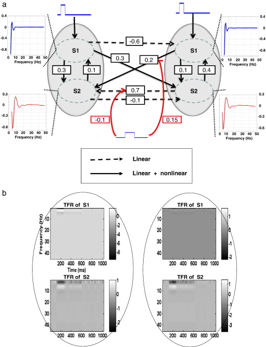
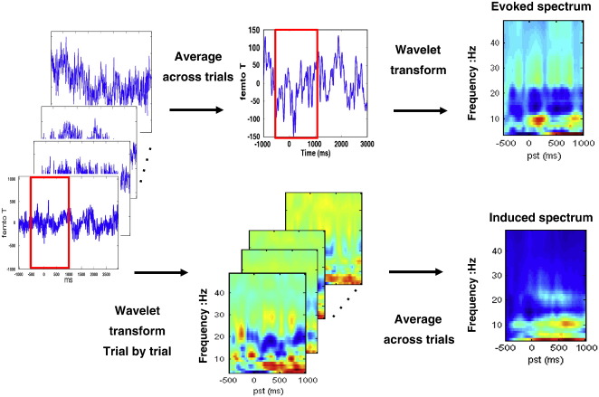
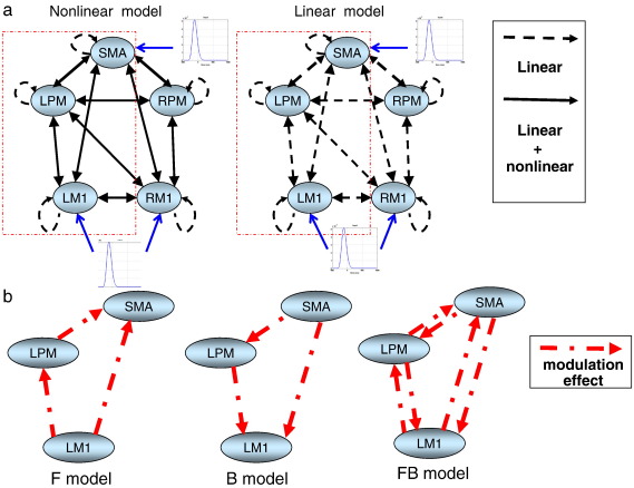
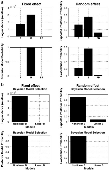
Neuronal responses exhibit two stimulus or task-related components: evoked and induced. The functional role of induced responses has been ascribed to 'top-down' modulation through backward connections and lateral interactions; as opposed to the bottom-up driving processes that may predominate in evoked components. The implication is that evoked and induced components may reflect different neuronal processes. The conventional way of separating evoked and induced responses assumes that they can be decomposed linearly; in that induced responses are the average of the power minus the power of the average (the evoked component). However, this decomposition may not hold if both components are generated by nonlinear processes. In this work, we propose a Dynamic Causal Model that models evoked and induced responses at the same time. This allows us to explain both components in terms of shared mechanisms (coupling) and changes in coupling that are necessary to explain any induced components. To establish the face validity of our approach, we used Bayesian Model Selection to show that the scheme can disambiguate between models of synthetic data that did and did not contain induced components. We then repeated the analysis using MEG data during a hand grip task to ask whether induced responses in motor control circuits are mediated by 'top-down' or backward connections. Our result provides empirical evidence that induced responses are more likely to reflect backward message passing in the brain, while evoked and induced components share certain characteristics and mechanisms.


Figures

Fig. 1
Fig. 1
(a) Simulation architecture and parameters. The two grey circles represent the two areas in this model, while S1 and S2 denote two frequency modes within each area. A frequency mode corresponds to a pattern of frequency-specific deviations from the baseline spectral profile (these patterns are shown as a function of frequency in the inserts). Time-dependent modulations of these frequency modes correspond to evoked and induced responses. The solid lines represent nonlinear connections because they connect different frequency modes, while the dashed lines couple the same frequency modes and therefore model linear coupling. The red lines indicate the connections that can change in a condition-specific fashion (here, whether we are trying to explain induced or purely evoked spectral responses). (b) The time-frequency data generated by this model. These are linear mixtures of the two time-varying frequency modes above. Left column: Area 1; right column: Area 2.
Fig. 2
Fig. 2
The flowchart of data preparation for evoked (upper) and induced (lower) responses. The red rectangles represent the time window of interest from − 500 to 1000 ms. Note that the spectral densities have been normalised individually.
Fig. 3
Fig. 3
Model specifications of Forward (F), Backward (B), and Forward-Backward (FB) models based on the previous MEG study (Chen et al., 2010). (a) The basic network configuration has a left hemispheric dominance and extrinsic nonlinear connections (left) and an ‘all-linear’ model (right) is included to test whether nonlinear mechanism is crucial to explain the dataset, in particular, the induced responses. The blue arrows specify the areas which receive the exogenous input perturbation. The exogenous input was modelled using a gamma function with two normally distributed parameters estimated from the data. (b) The modulation effects are allowed in the left hemisphere (red rectangle) in only forward (F model), or backward (B model) or both forward and backward connections (FB model).
Fig. 4
Fig. 4
(a) Bayesian Model Selection results of nonlinear models at the group level under fixed effects (left) and random effects (right) assumptions suggest that the B (Backward) model is the winning model among the models tested. (b) Bayesian Model Selection between linear and nonlinear B models shows that nonlinear B model is superior to the linear B model and supported the idea that nonlinear connectivity is essential to the generating of induced responses.
Fig. 5
Fig. 5
The corresponding SPMs (T-statistic map; thresholded at p < 0.005 uncorrected) of the connection-specific modulation matrices for significant ‘excitatory’ (positive; red blobs) and ‘inhibitory’ (negative; green blobs) effects respectively. The yellow arrows indicate several instances of consistent nonlinear interactions in the backward connections across all subjects.

References

    1. Angelucci A., Levitt J.B., Walton E.J., Hupe J.M., Bullier J., Lund J.S. Circuits for local and global signal integration in primary visual cortex. J. Neurosci. 2002;22:8633–8646. - PMC - PubMed
    1. Angelucci A., Levitt J.B., Lund J.S. Anatomical origins of the classical receptive field and modulatory surround field of single neurons in macaque visual cortical area V1. Prog. Brain Res. 2002;136:373–388. - PubMed
    1. Aoki F., Fetz E.E., Shupe L., Lettich E., Ojemann G.A. Changes in power and coherence of brain activity in human sensorimotor cortex during performance of visuomotor tasks. Biosystems. 2001;63:89–99. - PubMed
    1. Box G.E.P., Draper N.R. John Wiley and Sons; New York: 1987. Empirical Model-Building and Response Surfaces.
    1. Chen C.C., Kiebel S.J., Friston K.J. Dynamic causal modelling of induced responses. NeuroImage. 2008;41:1293. - PubMed

Publication types