

Capsule endoscopy in nonresponsive celiac disease
- PMID: 21835400
- PMCID: PMC3499038
- DOI: 10.1016/j.gie.2011.05.049
Capsule endoscopy in nonresponsive celiac disease
Abstract
Background: Nonresponsive celiac disease (CD) is defined by persistent or recurrent symptoms, common after treatment with a gluten-free diet (GFD).
Objective: To evaluate the utility of capsule endoscopy (CE) in nonresponsive CD.
Design: Case-control study.
Setting: Tertiary-care center.
Patients: Forty-two consecutive patients with nonresponsive CD and 84 age- and sex-matched CD-free controls who underwent CE were included. In addition, capsules taken after treatment with a GFD were retrospectively evaluated in 30 patients with uncomplicated CD.
Intervention: CE.
Main outcome measurements: Diagnostic accuracy of CE for the detection of mucosal abnormalities in nonresponsive CD.
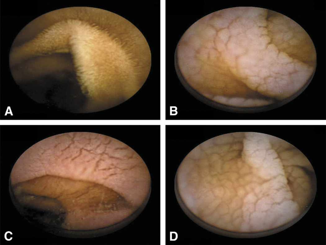
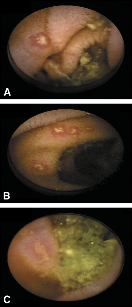
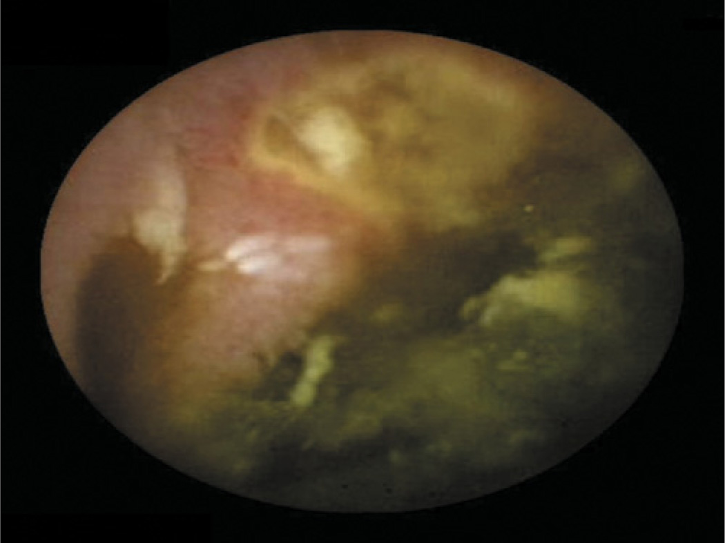
Results: Macroscopic features of villous atrophy were detected in 13 of 42 patients (31%) with nonresponsive CD compared with none among 84 CD-free controls and 14 of 30 patients (47%) with uncomplicated CD. Among nonresponsive CD cases, the overall sensitivity and specificity of CE for the detection of any degree of villous atrophy as graded by histology were 56% and 85%, respectively. Single or multiple erosions/ulcerations of the gut were observed in 19% of nonresponsive CD patients, 18% of CD-free controls, and 31% of patients with uncomplicated CD (P = .35). The presence of erosions/ulcerations was associated with increased aspirin/nonsteroidal anti-inflammatory drug use in nonresponsive CD (P =.05). Two severe complications (ulcerative jejunitis and adenocarcinoma) were detected by CE in nonresponsive CD.
Limitations: Single-center, retrospective study.
Conclusions: Mucosal abnormalities were observed by CE in patients with both nonresponsive CD and uncomplicated CD. CE can detect severe complications in patients with nonresponsive CD.
Copyright © 2011 American Society for Gastrointestinal Endoscopy. Published by Mosby, Inc. All rights reserved.
Figures
Comment in
-
The role of capsule endoscopy in patients with nonresponsive celiac disease.Gastrointest Endosc. 2011 Dec;74(6):1323-4. doi: 10.1016/j.gie.2011.07.021. Gastrointest Endosc. 2011. PMID: 22136777 No abstract available.
Similar articles
-
Capsule endoscopy in adult celiac disease: a potential role in equivocal cases of celiac disease?Gastrointest Endosc. 2013 Feb;77(2):227-32. doi: 10.1016/j.gie.2012.09.031. Epub 2012 Nov 30. Gastrointest Endosc. 2013. PMID: 23200728 Clinical Trial.
-
The role of capsule endoscopy in patients with nonresponsive celiac disease.Gastrointest Endosc. 2011 Dec;74(6):1323-4. doi: 10.1016/j.gie.2011.07.021. Gastrointest Endosc. 2011. PMID: 22136777 No abstract available.
-
Role of capsule endoscopy in alarm features and non-responsive celiac disease: A European multicenter study.Dig Endosc. 2018 Jul;30(4):461-466. doi: 10.1111/den.13002. Epub 2018 Jan 22. Dig Endosc. 2018. PMID: 29253321
-
Monitoring nonresponsive patients who have celiac disease.Gastrointest Endosc Clin N Am. 2006 Apr;16(2):317-27. doi: 10.1016/j.giec.2006.03.005. Gastrointest Endosc Clin N Am. 2006. PMID: 16644460 Review.
-
Tests for Serum Transglutaminase and Endomysial Antibodies Do Not Detect Most Patients With Celiac Disease and Persistent Villous Atrophy on Gluten-free Diets: a Meta-analysis.Gastroenterology. 2017 Sep;153(3):689-701.e1. doi: 10.1053/j.gastro.2017.05.015. Epub 2017 May 22. Gastroenterology. 2017. PMID: 28545781 Free PMC article. Review.
Cited by
-
Small bowel capsule endoscopy: Where are we after almost 15 years of use?World J Gastrointest Endosc. 2015 Jan 16;7(1):13-36. doi: 10.4253/wjge.v7.i1.13. World J Gastrointest Endosc. 2015. PMID: 25610531 Free PMC article. Review.
-
Diagnosis and management of adult coeliac disease: guidelines from the British Society of Gastroenterology.Gut. 2014 Aug;63(8):1210-28. doi: 10.1136/gutjnl-2013-306578. Epub 2014 Jun 10. Gut. 2014. PMID: 24917550 Free PMC article.
-
Evaluating Responses to Gluten Challenge: A Randomized, Double-Blind, 2-Dose Gluten Challenge Trial.Gastroenterology. 2021 Feb;160(3):720-733.e8. doi: 10.1053/j.gastro.2020.10.040. Epub 2020 Oct 29. Gastroenterology. 2021. PMID: 33130104 Free PMC article. Clinical Trial.
-
Optimising the use of small bowel endoscopy: a practical guide.Frontline Gastroenterol. 2019 Apr;10(2):171-176. doi: 10.1136/flgastro-2018-101077. Epub 2019 Jan 4. Frontline Gastroenterol. 2019. PMID: 31205659 Free PMC article. Review.
-
Capsule endoscopy: current practice and future directions.World J Gastroenterol. 2014 Jun 28;20(24):7752-9. doi: 10.3748/wjg.v20.i24.7752. World J Gastroenterol. 2014. PMID: 24976712 Free PMC article. Review.
References
-
- Di Sabatino A, Corazza GR. Coeliac disease. Lancet. 2009;373:1480–1493. - PubMed
-
- Rostom A, Murray JA, Kagnoff MF. American Gastroenterological Association (AGA) Institute technical review on the diagnosis and management of celiac disease. Gastroenterology. 2006;131:1981–2002. - PubMed
-
- Abdulkarim AS, Burgart LJ, See J, et al. Etiology of nonresponsive celiac disease: results of a systematic approach. Am J Gastroenterol. 2002;97:2016–2021. - PubMed
-
- Leffler DA, Dennis M, Hyett B, et al. Etiologies and predictors of diagnosis in nonresponsive celiac disease. Clin Gastroenterol Hepatol. 2007;5:445–450. - PubMed
-
- Biagi F, Corazza GR. Defining gluten refractory enteropathy. Eur J Gastroenterol Hepatol. 2001;13:561–565. - PubMed
Publication types
MeSH terms
Grants and funding
LinkOut - more resources
Full Text Sources
Medical
