. 2011 Nov;219(1):33-9.
doi: 10.1016/j.atherosclerosis.2011.07.012. Epub 2011 Jul 20.

Perivascular visceral adipose tissue induces atherosclerosis in apolipoprotein E deficient mice

Affiliations

Perivascular visceral adipose tissue induces atherosclerosis in apolipoprotein E deficient mice

M K Öhman et al. Atherosclerosis. 2011 Nov.

Abstract

Objective: Epicardial adipose tissue is associated with coronary artery disease, however the causal relationship between perivascular adipose tissue and local atherogenesis is unclear.

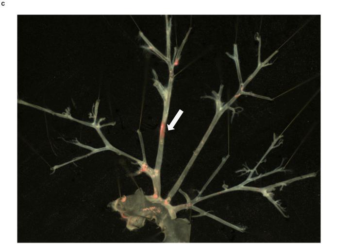
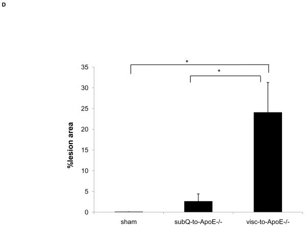
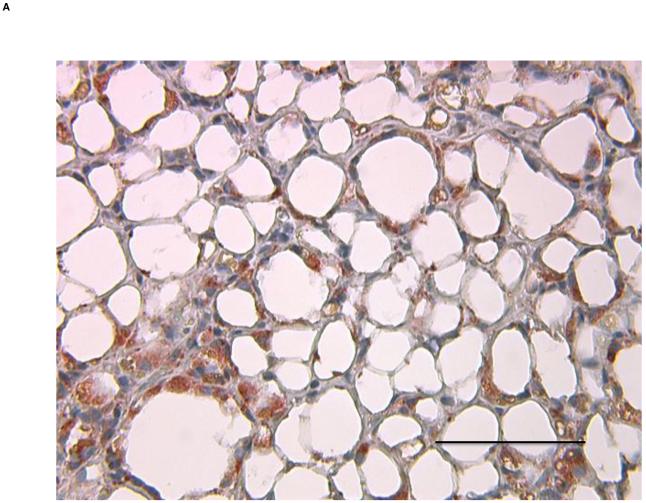
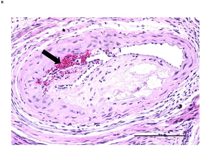
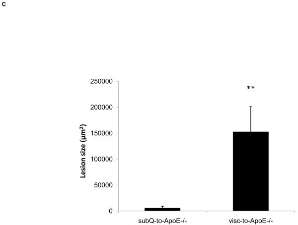
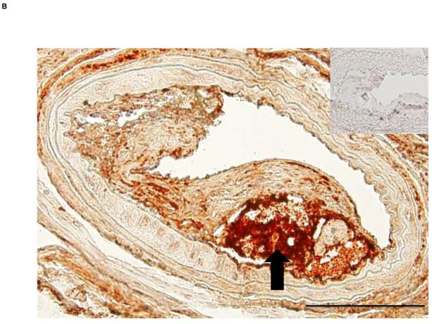
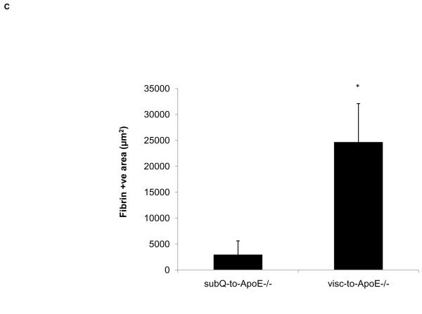
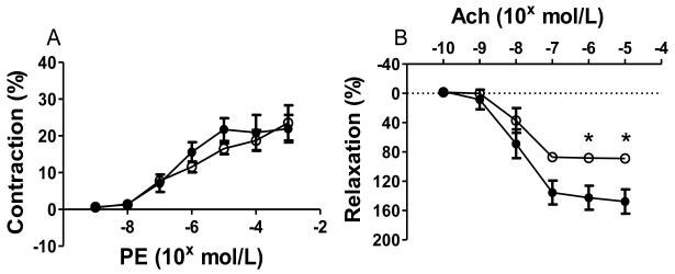
Methods and results: Apolipoprotein E deficient (ApoE(-/-)) mice underwent transplantation of visceral or subcutaneous adipose tissue immediately adjacent to the right common carotid artery. Carotid arteries with fat transplants were analyzed for atherosclerosis by surface oil-red-O staining and cross-sectional analysis. Vascular function of the carotid arteries was assessed using pressure myography. Visceral fat transplants were also performed to ApoE(-/-) mice with neutralization of P-selectin glycoprotein ligand-1 (Psgl-1). Atherosclerosis surface area and lesion thickness were greater in mice receiving the perivascular visceral fat compared to the subcutaneous fat. Mice with visceral fat transplants also displayed more complicated atherosclerotic lesions with evidence of atherothrombosis. Serum Mcp-1 was higher in mice receiving visceral fat transplants compared to subcutaneous transplants. Visceral fat transplantation also caused impaired endothelial-dependent relaxation of the carotid artery. Psgl-1 deficiency or neutralization of Psgl-1 with an anti-Psgl-1 antibody was protective against perivascular visceral adipose tissue-induced atherosclerosis and was associated with reduced Mcp-1 levels.

Conclusions: Perivascular visceral fat leads to endothelial dysfunction and accelerated atherosclerosis. This proatherogenic effect of perivascular adipose tissue is blocked by neutralization of Psgl-1.


Figures

Figure 1
Figure 1. Local atherosclerosis increased in mice with perivascular visceral fat transplantation
Oil-red-o stained aortic arch and major branches of ApoE−/− mouse transplanted with A) visceral (visc-to-ApoE−/−) adipose tissue, B) sham operated ApoE−/− mouse and C) ApoE−/− mouse transplanted with subcutaneous (subQ-to-ApoE−/−) adipose tissue. D) Lesion surface area in operated ApoE−/− mice. *p<0.05. Arrows point to the site of fat transplantation on the right common carotid arteries.
Figure 2
Figure 2. Inflammatory infiltrate in transplanted adipose tissue
A) Transplanted visceral adipose tissue and B) endogenous visceral adipose tissue stained with Mac3 antibody. Magnification 40x, scale bar =100 μm.
Figure 2
Figure 2. Inflammatory infiltrate in transplanted adipose tissue
A) Transplanted visceral adipose tissue and B) endogenous visceral adipose tissue stained with Mac3 antibody. Magnification 40x, scale bar =100 μm.
Figure 3
Figure 3. Perivascular visceral adipose tissue increases lesion thickness
H&E stained cross sections of the right common carotid artery from ApoE−/− mice with A) subcutaneous and B) visceral adipose tissue transplant. Arrow pointing to a potential intraplaque hemorrhage. Scale bar = 100 μm, magnification 40x. C) Lesion size in operated ApoE−/− mice. **p<0.01.
Figure 4
Figure 4. ApoE−/− mice transplanted with visceral fat display complicated lesions
A) Cross section of carotid artery stained with fibrin(ogen) antibody from ApoE−/− mice transplanted with A) subcutaneous and B) visceral fat. Inset shows negative control without primary antibody. Arrow points to necrotic core of the lesion. Scale bar = 100 μm, magnification 40x. C) Fibrin-positive area in lesions in operated mice. *p<0.05.
Figure 5
Figure 5. Perivascular visceral fat transplant leads to endothelial dysfunction
A) Vasoconstriction responses to phenylephrine (PE). B) Relaxation responses to acetylcholine (Ach). ApoE−/− mice with visceral (○) and subcutaneous (●) fat transplants. *p<0.05.

References

    1. See, R., Abdullah SM, McGuire DK, Khera A, Patel MJ, Lindsey JB, Grundy SM, de Lemos JA. The association of differing measures of overweight and obesity with prevalent atherosclerosis: The dallas heart study. Journal of the American College of Cardiology. 2007;50:752–759.

    1. Berg AH, Scherer PE. Adipose tissue, inflammation, and cardiovascular disease. Circ Res. 2005;96:939–949. - PubMed
    1. Konishi M, Sugiyama S, Sugamura K, Nozaki T, Ohba K, Matsubara J, Matsuzawa Y, Sumida H, Nagayoshi Y, Nakaura T, Awai K, Yamashita Y, Jinnouchi H, Matsui K, Kimura K, Umemura S, Ogawa H. Association of pericardial fat accumulation rather than abdominal obesity with coronary atherosclerotic plaque formation in patients with suspected coronary artery disease. Atherosclerosis. 2010;209:573–578. - PubMed
    1. Alexopoulos N, McLean DS, Janik M, Arepalli CD, Stillman AE, Raggi P. Epicardial adipose tissue and coronary artery plaque characteristics. Atherosclerosis. 2010;210:150–154. - PubMed
    1. Ueno K, Anzai T, Jinzaki M, Yamada M, Jo Y, Maekawa Y, Kawamura A, Yoshikawa T, Tanami Y, Sato K, Kuribayashi S, Ogawa S. Increased epicardial fat volume quantified by 64-multidetector computed tomography is associated with coronary atherosclerosis and totally occlusive lesions. Circulation Journal. 2009;73:1927–1933. - PubMed

Publication types