

Diffusion weighted imaging in predicting progression free survival in patients with squamous cell carcinomas of the head and neck treated with induction chemotherapy
- PMID: 21835649
- PMCID: PMC3168957
- DOI: 10.1016/j.acra.2011.06.009
Diffusion weighted imaging in predicting progression free survival in patients with squamous cell carcinomas of the head and neck treated with induction chemotherapy
Abstract
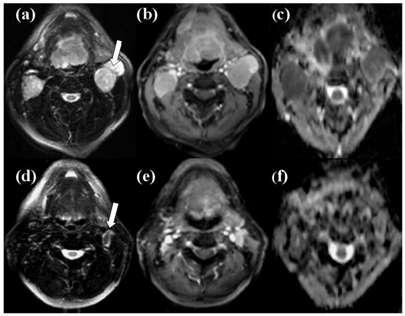
Rationale and objectives: The aim of this study was to assess the role of diffusion-weighted imaging in predicting progression-free survival in patients with head and neck squamous cell carcinoma (HNSCC) treated with induction chemotherapy.
Materials and methods: Eighteen patients with HNSCC underwent diffusion-weighted imaging studies prior to treatment and within 3 weeks after completion of induction chemotherapy. Median apparent diffusion coefficient (ADC) values were computed from the largest cervical metastatic lymph node. Percentage changes in ADC values from pretreatment to posttreatment time points were compared between alive and dead patients using the Mann-Whitney U test. P values < .05 were considered statistically significant.
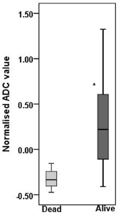
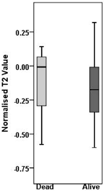
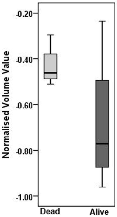
Results: A 22% increase in ADC was observed after induction chemotherapy in alive patients (n = 15), while patients who died from HNSCC (n = 3) demonstrated a 33% decrease in ADC. The difference in percentage change in ADC between alive and dead patients was significant (P = .039).
Conclusions: ADC may be a useful marker in predicting progression-free survival in patients with HNSCC undergoing induction chemotherapy.
Copyright © 2011 AUR. Published by Elsevier Inc. All rights reserved.
Figures
References
-
- Posner M, Vermorken JB. Induction therapy in the modern era of combined-modality therapy for locally advanced head and neck cancer. Semin Oncol. 2008;35(3):221–8. - PubMed
-
- Specenier PM, Vermorken JB. Neoadjuvant chemotherapy in head and neck cancer: should it be revisited? Cancer Lett. 2007;256(2):166–77. - PubMed
-
- Ensley JF, Jacobs JR, Weaver A, et al. Correlation between response to cisplatinum-combination chemotherapy and subsequent radiotherapy in previously untreated patients with advanced squamous cell cancers of the head and neck. Cancer. 1984;54(5):811–4. - PubMed
-
- Lee WC, Chavez YE, Baker T, Luce BR. Economic burden of heart failure: a summary of recent literature. Heart Lung. 2004;33(6):362–71. - PubMed
-
- Fury MG, Shah JP. Induction chemotherapy in the management of head and neck cancer. J Surg Oncol. 101(4):292–8. - PubMed
Publication types
MeSH terms
Substances
Grants and funding
LinkOut - more resources
Full Text Sources
Medical
