. 2011 Sep 2;43(5):738-50.
doi: 10.1016/j.molcel.2011.07.020. Epub 2011 Aug 11.

The mechanism of tail-anchored protein insertion into the ER membrane

Affiliations

The mechanism of tail-anchored protein insertion into the ER membrane

Fei Wang et al. Mol Cell. .

Abstract

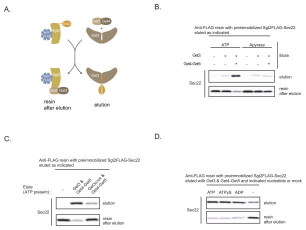
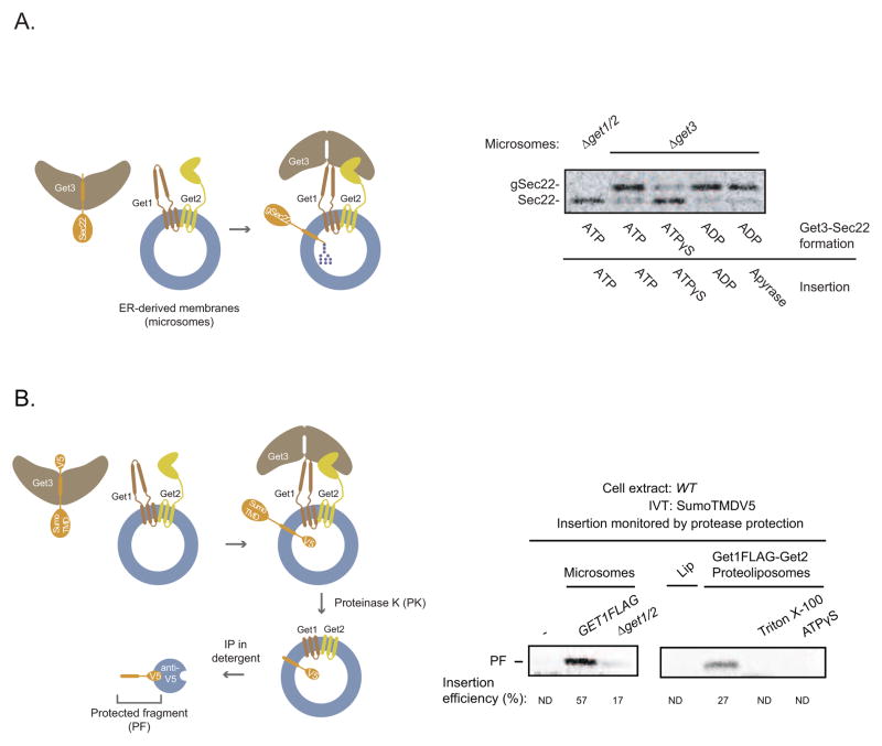
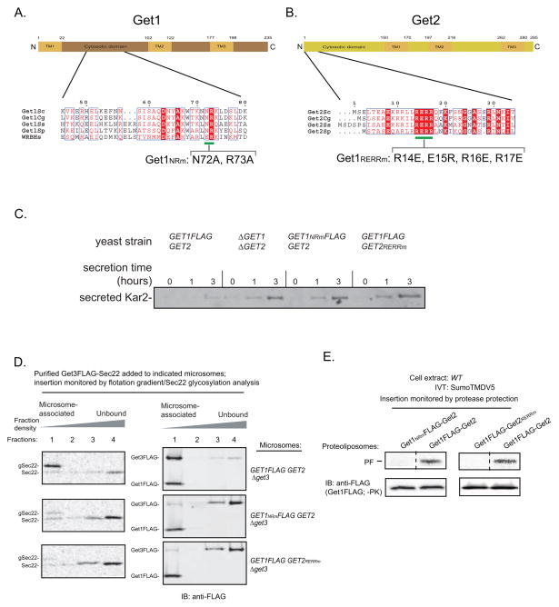
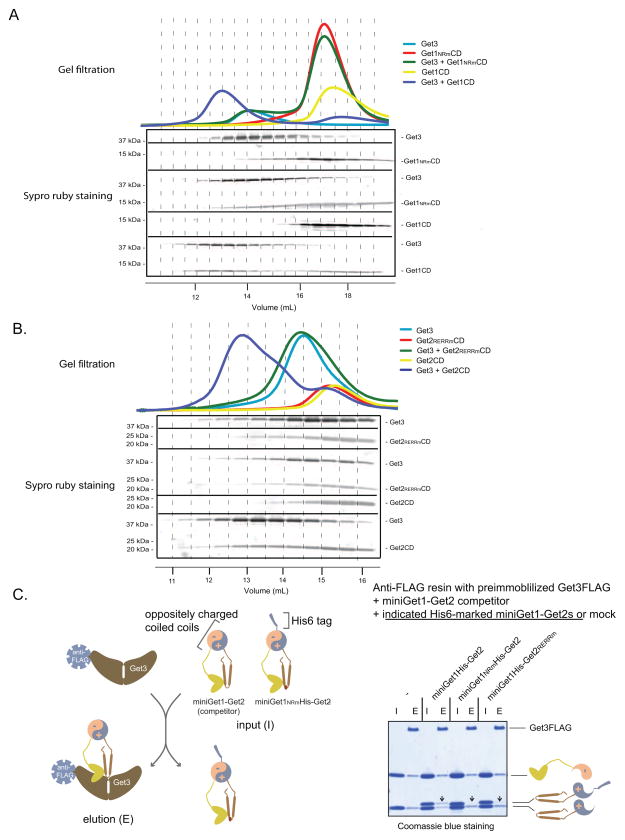
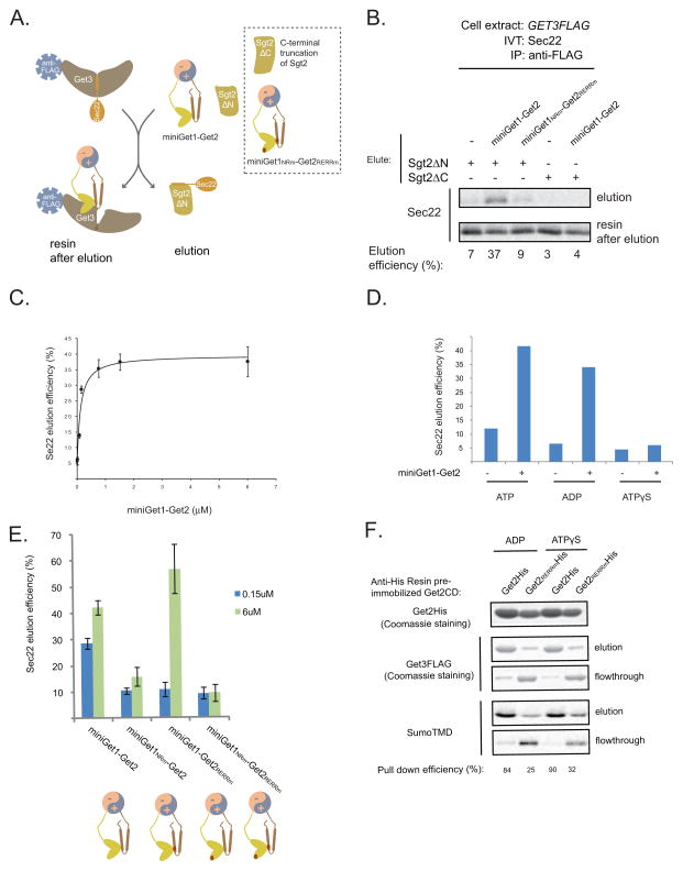
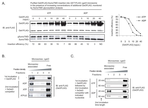
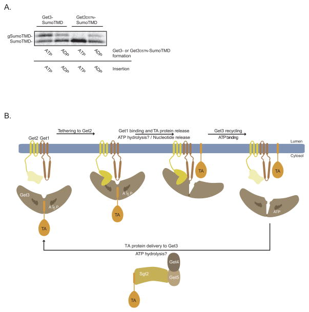
Tail-anchored (TA) proteins access the secretory pathway via posttranslational insertion of their C-terminal transmembrane domain into the endoplasmic reticulum (ER). Get3 is an ATPase that delivers TA proteins to the ER by interacting with the Get1-Get2 transmembrane complex, but how Get3's nucleotide cycle drives TA protein insertion remains unclear. Here, we establish that nucleotide binding to Get3 promotes Get3-TA protein complex formation by recruiting Get3 to a chaperone that hands over TA proteins to Get3. Biochemical reconstitution and mutagenesis reveal that the Get1-Get2 complex comprises the minimal TA protein insertion machinery with functionally critical cytosolic regions. By engineering a soluble heterodimer of Get1-Get2 cytosolic domains, we uncover the mechanism of TA protein release from Get3: Get2 tethers Get3-TA protein complexes into proximity with the ATPase-dependent, substrate-releasing activity of Get1. Lastly, we show that ATP enhances Get3 dissociation from the membrane, thus freeing Get1-Get2 for new rounds of substrate insertion.


Figures

Figure 1
Figure 1. ATP Binding to Get3 Stimulates TA Protein Transfer from Sgt2-Get5-Get4 to Get3
(A) Schematic of the in vitro assay for studying TA protein handoff from Sgt2 to Get3. Sec22 is a SNARE TA protein. Sgt2-Sec22 immobilized on anti-FLAG resin comes from immunoprecipitation of the SGT2FLAG Δget3/5 extract with in vitro translated Sec22-encoding mRNA. Sgt2FLAG purified in the absence of Get5 lacks Get4, (see Wang et al., 2010 for more details). (B) In vitro translation of Sec22-encoding mRNA in the SGT2FLAG Δget3/5 extract in the presence of 35S-labeled methionine was followed by anti-FLAG immunoprecipitation (IP) and elution with the indicated Get proteins (Get3: 64 ng/μL; Get4-Get5: 96 ng/μL) or mock treatment for 20 minutes at room temperature. ATP (4 mM) or Apyrase (1 unit/μL) was also included during elution, as indicated. Following centrifugation, elutions were collected, the resin washed, and eluted again, but this time with gel loading buffer (resin after elution). The elutions were resolved by SDS-PAGE and analyzed by autoradiography. (C) Sec22 elution from preimmobilized Sgt2-Sec22 with either Get3 or Get3G30R in combination with Get4-Get5 or mock treatment was carried out as in (B). ATP (4 mM) was included in all elutions. (D) Sec22 elution from preimmobilized Sgt2-Sec22 with Get3 and Get4-Get5 in the presence of the indicated nucleotides (4 mM) or mock nucleotide treatment was carried out as in (B).
Figure 2
Figure 2. The Get1-Get2 Transmembrane Complex is the Minimal ER Membrane Machinery for TA Protein insertions
(A) (ON THE LEFT) Schematic showing glycosylation upon microsomal insertion of Sec22, which contains a carboxyl-terminal Opsin tag with an N-glycan acceptor site. (ON THE RIGHT) Sec22 eluted with Get3His and Get4-Get5 in the presence of the indicated nucleotides (3 mM) as in Figure 1D was immunoprecipitated with anti-His resin, washed, and eluted with imidazole (Figure S2A). Elutions were incubated with Δget3 microsomes (to eliminate any endogenous Get3 that remains stably bound to Get1-Get2 during microsome preparation) or Δget1/2 microsomes for 30 minutes at room temperature in the presence of the indicated nucleotides (4 mM) or Apyrase (5 units/μL). Samples were resolved by SDS-PAGE and analyzed by autoradiography. The positions of Sec22 and glycosylated Sec22 (gSec22) are indicated. (B) (ON THE LEFT) Schematic of protease protection assay for monitoring TA protein insertion. In vitro synthesis of SumoTMD with a C-terminal V5 epitope was followed by incubation with membranes containing Get1-Get2 and then treatment with proteinase K (PK). Following PK inactivation, digested membranes were solubilized with detergent (1% Triton) and subjected to immunoprecipitation (IP) with anti-V5 resin to detect the protected transmembrane domain fragment (PF) (see Figure S2C). (ON THE RIGHT) Wild-type (WT) extract with in vitro translated (IVT) SumoTMDV5-encoding mRNA was incubated with the indicated microsomes, liposomes (Lip), Get1FLAG-Get2 proteoliposomes, or mock incubated for 30 min at room temperature. Samples were then treated with proteinase K (PK) treatment, resolved by SDS-PAGE, and analyzed by autoradiography. Where indicated, ATPγS (3mM) and Triton X-100 (1%) were included at the time of proteoliposome or PK addition, respectively. Insertion efficiency is defined as the percentage of the protected fragment (PF) signal relative to the full-length signal prior to PK addition (not shown). Both signals were normalized for their methionine content. ND: not detected. Note that the version of SumoTMDV5 used here has a mutation in the N-glycan acceptor site to allow for direct comparison of the PF between microsomes (which would otherwise have caused the PF to become glycosylated) and proteoliposomes.
Figure 3
Figure 3. The Conserved Cytosolic Domains of Get1 and Get2 are Required to Recruit Get3-TA Protein Complexes for Insertion into the ER Membrane
(A) Schematic of Get1 topology with the residues bracketing the predicted transmembrane (TM) helices and the cytosolic domain indicated. Shown below is the 45–80 amino acid region of Saccharomyces cerevisiae Get1 aligned using Clustal W2 with Get1 homologs from Candida glabrata, Scheffersomyces stipitis, Schizosaccharomyces pombe, and the Homo sapiens tryptophan basic protein (WRB). ESPript 2.0 was used to highlight identical (white in color, boxed with red), well-conserved (red in color, boxed in white) residues. The green line indicates the conserved positions that were mutated to alanines in Get1NRm. (B) Schematic of Get2 topology with the residues bracketing the predicted transmembrane (TM) helices and the cytosolic domain indicated. Shown below is the 1–34 amino acid region of Saccharomyces cerevisiae Get2 aligned using Clustal W2 with several other Get2 fungal homologs described in (A). ESPript 2.0 was used to highlight residues as in (A). The green line indicates the conserved positions that were mutated to oppositely charged residues in Get2RERRm. (C) The indicated strains were grown to mid-log (OD600 0.6–0.8), washed, and shifted to fresh growth media at a starting OD600 ~0.5. At the indicated times, media samples were removed, TCA precipitated, and analyzed by SDS-PAGE and immunoblotting (IB) with the anti-Kar2 antibody. (D) In vitro translation of Sec22-encoding mRNA in the GET3FLAG extracts supplemented with additional 32 ng/μL Get3FLAG and 48 ng/μL Get4-Get5 to enhance Get3FLAG-Sec22 complex formation (data not shown). Following anti-FLAG immunoprecipitation (IP) and 3xFLAG peptide elution, elutions were split and incubated with the indicated microsomes for 30 minutes at room temperature. Samples were then overlayed with an Optiprep gradient and subjected to ultracentrifugation. Proteins were precipitated from each fraction and analyzed by autoradiography and immunoblotting (IB) with anti-FLAG. (E) Insertion of SumoTMDV5 into the indicated proteoliposomes was monitored by protease protection as in Figure 2B. Note that the proteoliposome samples were also analyzed prior to proteinase K (PK) treatment by SDS-PAGE analysis followed by immunoblotting (IB) with anti-FLAG antibody.
Figure 4
Figure 4. Cooperative Binding of Get3 to the Cytosolic Domains of Get1 and Get2
(A) and (B) The indicated proteins were analyzed by gel-filtration chromatography (Superdex 200 10/300 GL). The relevant fractions (indicated by dash lines connecting the top A280 traces with the elution volumes on the bottom) were resolved by SDS-PAGE and visualized by Sypro ruby staining (in the middle). (C) (ON THE LEFT) Schematic of miniGet1-Get2 binding competition assay illustrating that the miniGet1NRm-Get2 is outcompeted by the wild-type for binding to Get3 (see gel on the right). (ON THE RIGHT) Anti-FLAG resin with pre-immobilized Get3FLAG was incubated with the indicated His-marked miniGet1-Get2s and miniGet1-Get2 competitor inputs (I) (Get3FLAG:miniGet1His-Get2:miniGet1-Get2 molar ratio equals 1:2:2) in the presence of ADP (3 mM) for 20 minutes at room temperature. Following washing, the resin was eluted (E) with FLAG peptide. Samples were resolved by SDS-PAGE and visualized by Coomassie blue staining. Arrows point to the positions on the gel at which we expect to see His-tagged Get1 bands if they are able to compete for binding to Get3.
Figure 5
Figure 5. In Vitro Reconstitution Reveals a Dual Mechanism by which Get1 and Get2 Stimulate TA Protein Release from Get3
(A) Schematic showing release of Sec22 from immobilized Get3 by miniGet1-Get2. (B) In vitro translation (IVT) of Sec22-encoding mRNA in the GET3FLAG extract supplemented with additional recombinant Get3FLAG (32 ng/μL) to enhance Get3FLAG-Sec22 complex formation (data not shown). After anti-FLAG immunoprecipitation (IP), the washed resin was incubated with miniGet1-Get2s (6 μM), Sgt2ΔN (0.1μg/μL), and Sgt2ΔC (0.1 μg/μL), as indicated, for 20 minutes at room temperature. Following centrifugation, elutions were collected and the resin washed and eluted again, but this time with gel loading buffer (resin after elution). The elutions were resolved by SDS-PAGE and analyzed by autoradiography. Percentage of Sec22 eluted under these conditions (elution/[elution+resin after elution]x100%) is indicated at the bottom. (C) Dose-dependent elution of Sec22 from immobilized Get3 at different concentrations of miniGet1-Get2 (starting at 0.01 μM) in the presence of Sgt2ΔN (0.1 μg/μL). Samples were prepared and analyzed as in part (B). Average Sec22 elution efficiency and standard deviation from two independent experiments are plotted as a function of miniGet1-Get2 concentration. (D) Sec22 elution from immobilized Get3 in the absence or presence of miniGet1-Get2 (6 μM) and Sgt2ΔN (0.1 μg/μL) with the indicated nucleotides (2.75mM) present. Samples were prepared and analyzed as in part (B). (E) Sec22 elution from immobilized Get3FLAG-Sec22 by the indicated miniGet1-Get2s (0.15 or 6 μM) in the presence of ATP (2.75 mM). Elution was carried out in the presence of Sgt2ΔN (0.1 μg/μL) and analyzed as in Figure 5B. Average Sec22 elution efficiency and standard deviation from two independent experiments are plotted. (F) Get3FLAG-SumoTMD was purified as described in Figure 2B and added along with additional Get3FLAG (4 μg; this reduces non-specific binding of Get3FLAG-SumoTMD to the resin, data not shown) to Ni-NTA agarose resin with preimmobilized His-tagged versions of the Get2 or Get2RERRm cytosolic domains (12 μg) for 20 min at room temperature. Following centrifugation, the flowthrough was collected and the resin was washed and then eluted with SDS gel loading buffer. Samples were resolved by SDS-PAGE and visualized by Coomassie blue staining and autoradiography.
Figure 6
Figure 6. ATP Stimulates TA Protein Insertion when Get3 Dissociation From the Membrane is Rate-Limiting
(A) Get3FLAG-SumoTMD was purified as in Figure 3D and incubated with the GET1FLAG Δget3 microsomes in the presence of either ADP or ATP and additional Get3FLAG, as indicated, for 30 minutes at room temperature. Samples were resolved by SDS-PAGE and analyzed by autoradiography and immunoblotting (IB) with anti-FLAG. Insertion efficiency is defined as the percentage of SumoTMD that is glycosylated (gSumoTMD/[gSumoTMD+SumoTMD]x100%). The insertion efficiencies in the presence of ADP and ATP at different concentrations of added Get3FLAG is plotted on the right. ND: not detected. (B) Δget3 microsomes (0.8 A280 units) were incubated with Get3FLAG (0.1 μg) in the absence of nucleotide for 20 minutes at room temperature (1st incubation), and then split and incubated with the indicated nucleotides (4 mM) and Get3 competitor (0.5 μg; lacking the FLAG tag) for another 20 minutes at room temperature (2nd incubation). Samples were analyzed by flotation analysis and immunoblotting (IB) with anti-FLAG as described in Figure 3D. (C) Δget3 microsomes (0.8 A280 units) were incubated with Get3FLAG (0.1 μg) in the absence of nucleotide for 20 minutes at room temperature (1st incubation), and then split and incubated with the indicated nucleotides (4 mM) and Get3 competitor (0.5 μg; lacking the FLAG tag) for 20 or 80 minutes at room temperature (2nd incubation). Samples were analyzed by flotation analysis and immunoblotting (IB) with anti-FLAG as described in Figure 3D.
Figure 7
Figure 7. The ATPase Activity of Get3 Switches the GET Pathway from the Targeting to the Insertion Stage
A) Get3FLAG-SumoTMD and Get3D57NFLAG-SumoTMD complexes were purified from wild-type yeast cell extract as in Figure 3D with the following modification. Complexes prepared in the presence of ADP were first passed through G-50 spin columns after in vitro translation to remove nucleotides before adding ADP and recombinant proteins as in Figure 3D. Get3FLAG-SumoTMD and Get3FLAG D57N-SumoTMD were incubated with the GET1FLAG Δget3 microsomes in the presence of either ADP or ATP, as indicated, for 30 minutes at room temperature. Samples were resolved by SDS-PAGE and analyzed by autoradiography as in Figure 6A. B) Schematic showing the role of nucleotide and GET pathway components during TA protein targeting and insertion into the ER membrane. See Discussion for more details.

Similar articles

Cited by

References

    1. Auld K, Hitchcock A, Doherty H, Frietze S, Huang L, Silver P. The conserved ATPase Get3/Arr4 modulates the activity of membrane associated proteins in Saccharomyces cerevisiae. Genetics. 2006:215–227. - PMC - PubMed
    1. Borgese N, Fasana E. Targeting pathways of C-tail-anchored proteins. Biochim Biophys Acta. 2011;1808:937–946. - PubMed
    1. Bozkurt G, Stjepanovic G, Vilardi F, Amlacher S, Wild K, Bange G, Favaloro V, Rippe K, Hurt E, Dobberstein B, et al. Structural insights into tail-anchored protein binding and membrane insertion by Get3. Proc Natl Acad Sci U S A. 2009:21131–21136. - PMC - PubMed
    1. Chang Y, Chuang Y, Ho Y, Cheng M, Sun Y, Hsiao C, Wang C. Crystal structure of Get4/Get5 complex and its interactions with Sgt2, Get3 and Ydj1. J Biol Chem 2010 - PMC - PubMed
    1. Chartron JW, Suloway CJ, Zaslaver M, Clemons WM., Jr Structural characterization of the Get4/Get5 complex and its interaction with Get3. Proc Natl Acad Sci U S A. 2010;107:12127–12132. - PMC - PubMed

Publication types

MeSH terms

Substances