. 2011 Oct;85(20):10669-81.
doi: 10.1128/JVI.05249-11. Epub 2011 Aug 10.

Primary infection by a human immunodeficiency virus with atypical coreceptor tropism

Affiliations

Primary infection by a human immunodeficiency virus with atypical coreceptor tropism

Chunlai Jiang et al. J Virol. 2011 Oct.

Abstract

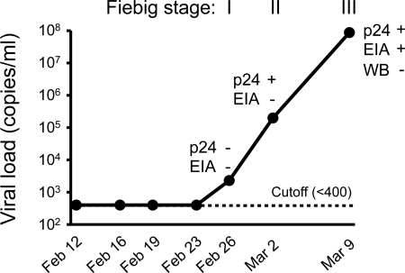
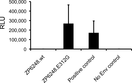
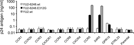
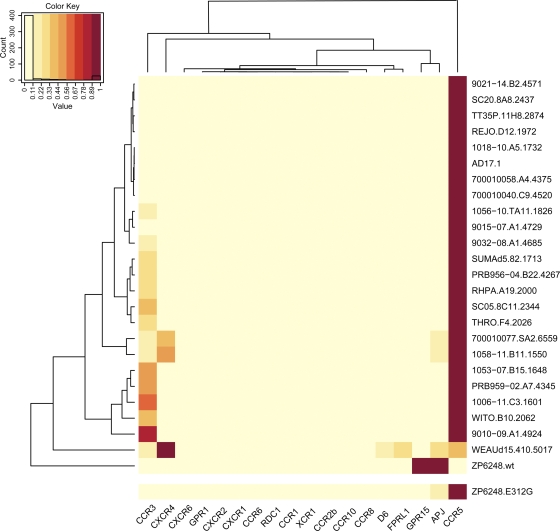
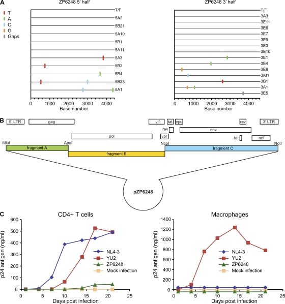
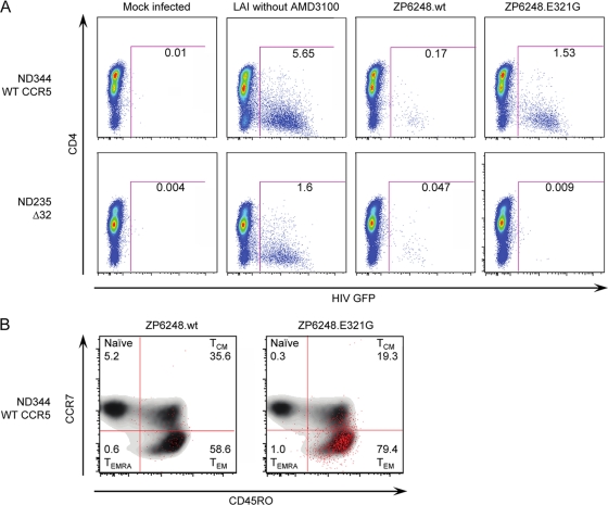
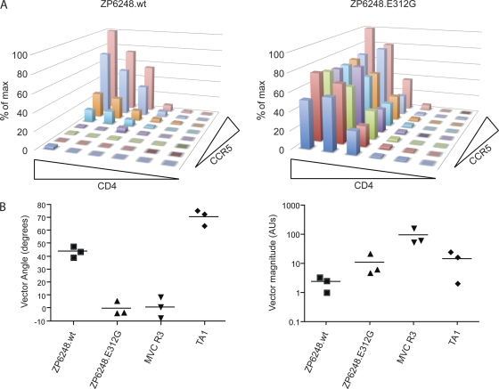
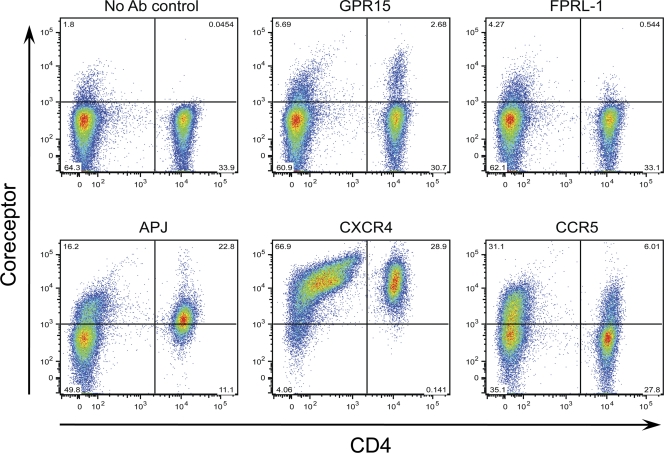
The great majority of human immunodeficiency virus type 1 (HIV-1) strains enter CD4+ target cells by interacting with one of two coreceptors, CCR5 or CXCR4. Here we describe a transmitted/founder (T/F) virus (ZP6248) that was profoundly impaired in its ability to utilize CCR5 and CXCR4 coreceptors on multiple CD4+ cell lines as well as primary human CD4+ T cells and macrophages in vitro yet replicated to very high titers (>80 million RNA copies/ml) in an acutely infected individual. Interestingly, the envelope (Env) glycoprotein of this clade B virus had a rare GPEK sequence in the crown of its third variable loop (V3) rather than the consensus GPGR sequence. Extensive sequencing of sequential plasma samples showed that the GPEK sequence was present in virtually all Envs, including those from the earliest time points after infection. The molecularly cloned (single) T/F virus was able to replicate, albeit poorly, in cells obtained from ccr5Δ32 homozygous donors. The ZP6248 T/F virus could also infect cell lines overexpressing the alternative coreceptors GPR15, APJ, and FPRL-1. A single mutation in the V3 crown sequence (GPEK->GPGK) of ZP6248 restored its infectivity in CCR5+ cells but reduced its ability to replicate in GPR15+ cells, indicating that the V3 crown motif played an important role in usage of this alternative coreceptor. These results suggest that the ZP6248 T/F virus established an acute in vivo infection by using coreceptor(s) other than CCR5 or CXCR4 or that the CCR5 coreceptor existed in an unusual conformation in this individual.


Figures

Fig. 1.
Fig. 1.
Laboratory staging of acute HIV-1 infection in subject ZP6248. Plasma viral load (RNA copies/ml) was determined using the COBAS Amplicor HIV-1 monitor test (the threshold of the assay is 400 copies/ml). p24 antigen was detected using the Alliance HIV-1 p24 Antigen ELISA Kit (PerkinElmer, Boston, MA). HIV-1 specific antibodies were detected using the GS HIV-1/HIV-2 plus O EIA kit and the GS HIV-1 Western blot kit (Bio-Rad, Hercules, CA). The temporal appearance of HIV-1 specific markers according to the classification by Fiebig (21) is shown.
Fig. 2.
Fig. 2.
Identification of a rare GPEK V3 crown sequence in the ZP6248 envelope glycoprotein. (A) Single genome amplification (SGA) was used to infer the transmitted founder (T/F) env sequence as described (28). A Highlighter plot denotes the location of nucleotide substitutions in each SGA-derived env sequence compared to the T/F virus. The position of these substitutions is indicated on the bottom (p16 contains G-to-A changes characteristic of APOBEC-mediated hypermutation). Nucleotide substitutions and gaps are color coded (all env sequences were derived from the 9 March time point). (B) Alignment of Env protein sequences in the V3 domain. Sequences from three time points (9 March, 2 March, and 26 February) are compared to the subtype B consensus sequence. The rare EK V3 crown motif found in all ZP6248 sequences is highlighted in red. Dashes in the alignment indicate sequence identity to the consensus; dots indicate deletions.
Fig. 3.
Fig. 3.
Infectivity of pseudoviruses containing ZP6248 Env glycoproteins. Viral pseudotypes bearing the indicated Env glycoproteins were used to infect TZM-bl cells stably expressing CD4, CXCR4, and CCR5. Infectivity was determined by measuring relative light units (RLU) in cell lysates 48 h after infection. ZP6248.wt represents the T/F Env, while ZP6248.E312G contained a single amino acid substitution in the V3 crown. A subtype C Env (ZM53 [32]) was used as a positive control, while Env-deficient pseudotypes (SG3Δenv) were used as a negative control.
Fig. 4.
Fig. 4.
Detection of the rare ZP6248.E321G mutation during acute infection. The presence of the ZP6248.E321G mutation (A to G) was examined by parallel allele-specific sequencing (PASS) analysis in plasma collected at the 9 March time point. In this assay, cDNA annealed to an acrydite-modified primer is immobilized in an acrylamide gel, after which in-gel PCR is performed, with the resulting products accumulating around the individual cDNA templates. Sequencing primers that anneal just upstream of the mutation site in V3 (GAA->GGA) can be used to distinguish wt and mutant bases at the same position by single-base extension using Cy3- and Cy5-labeled adenosine (wt) and guanosine (mutant), respectively. The gel was scanned to obtain images with a GenePix 4000B microarray scanner, and the spot number was counted using the Progenesis PG200 software. Green and red spots indicate wt and mutant bases, respectively, detected in individual viral genomes. Two mutants were identified by arrows. A partial image from one of the three experiments is shown.
Fig. 5.
Fig. 5.
Coreceptor usage of ZP6248 Env. GHOST (3) cell lines expressing CD4 and the indicated coreceptors were infected with two replication-competent YU2/ZP6248 chimeras (YU2-6248.wt and YU2-6248.E312G) as well as wild-type HIV-1 YU2.wt. Viruses normalized by p24 content (100 ng) were used to infect cells, and virus replication was monitored by measuring p24 antigen in culture supernatants 7 days after infection. The y axis shows the mean of p24 concentrations from three independent experiments. Error bars represent one standard error of the mean.
Fig. 6.
Fig. 6.
Comparison of the ZP6248 Env tropism to that of other subtype B T/F Envs. Entry of ZP6248.wt and ZP6248.E312G pseudotypes into 18 different NP-2 cell lines stably expressing CD4 and the indicated seven-transmembrane domain receptors was compared to that of 24 clade B T/F Envs. The cell line that was infected most efficiently for each virus was normalized to one (dark red), while the entry into the other cell lines was expressed as a fraction of this value as indicated by the key within the figure. Dendrograms on the top and left of the heat map represent hierarchical clustering of the data that show coreceptors with similar patterns of usage by Envs (top) and Envs with similar tropism for coreceptor usage (left).
Fig. 7.
Fig. 7.
Protein expression from transfected cells. 293T cells were transfected with the proviral clones indicated, and cell lysates and viral supernatants were harvested 48 h posttransfection. Protein expression and processing was examined by Western blot analysis using an HIV-1-positive human serum sample and a mouse MAb (13D5) specific for the HIV-1 Env protein (31). The position of the Env precursor (gp160) and the extracellular domain (gp120) are indicated. Protein standards are indicated on the left.
Fig. 8.
Fig. 8.
Molecular cloning and biological characterization of the T/F virus of subject ZP6248. (A) 5′ (4,377 bp) and 3′ (4,528 bp) half-genomes overlapping by 66 bp were amplified by SGA from plasma viral RNA (9 March time point) and used to infer the sequence of the single T/F virus infecting subject ZP6248 as previously described (22, 38, 54). The Highlighter plots denote the location of nucleotide substitutions compared to the inferred T/F sequence, with their position indicated on the bottom. Nucleotide substitutions and gaps are color coded. (B) The full-length ZP6248 genome was synthesized in three overlapping fragments and cloned into a modified pBR322 vector as described previously (59). (C) Viruses harvested from transfected 293T cells were examined for their ability to replicate in activated primary human CD4+ lymphocytes (left) and monocyte-derived macrophages (right) from the same donor. The x axis indicates days postinfection; the y axis denotes p24 antigen concentration (ng/ml) in the culture supernatant.
Fig. 9.
Fig. 9.
Infection of human CD4+ T cells. Env pseudovirions expressing a GFP reporter were used to infect primary ccr5 Δ32/Δ32 or ccr5 wt CD4+ T cells in the presence of the CXCR4 antagonist AMD3100. Viral infection was assessed by flow cytometry after gating on live CD3+ cell singlets. (A) ccr5 Δ32/Δ32 or ccr5 wt CD4+ T cells were infected with ZP6248.wt and ZP6248.E312G in the presence of AMD3100. Mock-infected cells were used as a negative control, while the X4 HIV-1 strain LAI was used as a positive control in the absence of AMD3100. (B) Infected cells were back-gated onto memory markers CCR7 and CD45R0 (CCR7+ CD45RO, naïve; CCR7+ CD45RO+, central memory; CCR7 CD45RO+ effector memory; CCR7 CD45RO, effector memory RA) to evaluate memory subset tropism of ZP6248.wt and ZP6248.E312G on CCR5 wild-type cells (ND344). The uninfected cells are shown in a contour plot to better reveal the distribution of infected (GFP+ cells) shown as red dots. 174 and 1,742 GFP+ events were collected for PZ6248.wt and ZP6248.E312G infected cells, respectively. The data are shown from one of five independent experiments with cells from five ccr5 wt donors and two ccr5Δ32 homozygous donors.
Fig. 10.
Fig. 10.
ZP6248.wt inefficiently utilizes CCR5. (A) ZP6248.wt and ZP6248.E312G pseudovirions were used to infect affinofile cells which express CD4 and CCR5 with independent induction systems. ZP6248.wt requires high CCR5 expression for infection compared to ZP6248.E312G, which can utilize very low levels of CCR5. Both Envs require moderate CD4 expression for infection. (B) The sensitivity to changes in CD4 and CCR5 levels was quantified by viral entry receptor sensitivity analysis (VERSA), which yields a single vector angle. Lower vector angles correspond to Envs that utilize CCR5 very efficiently as seen with the maraviroc-resistant Env MVC R3 (1°), while higher vector angles are associated with inefficient CCR5 use as seen with the V3-loop truncated virus TA1 (71°). ZP6248.wt has a vector angle of 44° compared to 0° for ZP6248.E312G. In addition, the vector magnitude can be quantified to assess overall infectivity. ZP6248.wt is 5-fold less infectious than ZP6248.E312G and 40-fold less infectious than MVC R3, consistent with other cell lines and primary cell data.
Fig. 11.
Fig. 11.
Coreceptor expression on peripheral blood mononuclear cells. PHA-stimulated or nonstimulated cells were labeled with antibodies specific for GPR15, FPRL-1, APJ, CXCR4, and CCR5, and the percentage of positive cells was determined. Representative results from one of five blood donors are shown. The x axis indicates CD4+ T cells, while the y axis denotes cells expressing the tested coreceptors. No significant differences for expression of GPR15, FPRL-1, and APJ were noticed on PHA-stimulated and nonstimulated cells, and the results from nonstimulated cells are shown.

Similar articles

Cited by

References

    1. Abrahams M. R., et al. 2009. Quantitating the multiplicity of infection with human immunodeficiency virus type 1 subtype C reveals a non-Poisson distribution of transmitted variants. J. Virol. 83:3556–3567 - PMC - PubMed
    1. Baba M., Miyake H., Wang X., Okamoto M., Takashima K. 2007. Isolation and characterization of human immunodeficiency virus type 1 resistant to the small-molecule CCR5 antagonist TAK-652. Antimicrob. Agents Chemother. 51:707–715 - PMC - PubMed
    1. Begaud E., et al. 2003. Broad spectrum of coreceptor usage and rapid disease progression in HIV-1-infected individuals from Central African Republic. AIDS Res. Hum. Retroviruses 19:551–560 - PubMed
    1. Berger E. A., Murphy P. M., Farber J. M. 1999. Chemokine receptors as HIV-1 coreceptors: roles in viral entry, tropism, and disease. Annu. Rev. Immunol. 17:657–700 - PubMed
    1. Berro R., et al. 2011. Multiple CCR5 conformations on the cell surface are used differentially by human immunodeficiency viruses resistant or sensitive to CCR5 inhibitors. J. Virol. 85:8227–8240 - PMC - PubMed

Publication types

MeSH terms