. 2011 Nov;85(21):11220-34.
doi: 10.1128/JVI.05238-11. Epub 2011 Aug 10.

Human and rhesus APOBEC3D, APOBEC3F, APOBEC3G, and APOBEC3H demonstrate a conserved capacity to restrict Vif-deficient HIV-1

Affiliations

Human and rhesus APOBEC3D, APOBEC3F, APOBEC3G, and APOBEC3H demonstrate a conserved capacity to restrict Vif-deficient HIV-1

Judd F Hultquist et al. J Virol. 2011 Nov.

Abstract

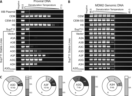
Successful intracellular pathogens must evade or neutralize the innate immune defenses of their host cells and render the cellular environment permissive for replication. For example, to replicate efficiently in CD4(+) T lymphocytes, human immunodeficiency virus type 1 (HIV-1) encodes a protein called viral infectivity factor (Vif) that promotes pathogenesis by triggering the degradation of the retrovirus restriction factor APOBEC3G. Other APOBEC3 proteins have been implicated in HIV-1 restriction, but the relevant repertoire remains ambiguous. Here we present the first comprehensive analysis of the complete, seven-member human and rhesus APOBEC3 families in HIV-1 restriction. In addition to APOBEC3G, we find that three other human APOBEC3 proteins, APOBEC3D, APOBEC3F, and APOBEC3H, are all potent HIV-1 restriction factors. These four proteins are expressed in CD4(+) T lymphocytes, are packaged into and restrict Vif-deficient HIV-1 when stably expressed in T cells, mutate proviral DNA, and are counteracted by HIV-1 Vif. Furthermore, APOBEC3D, APOBEC3F, APOBEC3G, and APOBEC3H of the rhesus macaque also are packaged into and restrict Vif-deficient HIV-1 when stably expressed in T cells, and they are all neutralized by the simian immunodeficiency virus Vif protein. On the other hand, neither human nor rhesus APOBEC3A, APOBEC3B, nor APOBEC3C had a significant impact on HIV-1 replication. These data strongly implicate a combination of four APOBEC3 proteins--APOBEC3D, APOBEC3F, APOBEC3G, and APOBEC3H--in HIV-1 restriction.


Figures

Fig. 1.
Fig. 1.
Human A3C, A3G, and A3H are induced in HIV-infected CD4+ T lymphocytes. (A) Histograms depicting the staining of PBMCs and naïve CD4+ T lymphocytes with a PE-conjugated antibody against CD4 after purification by negative selection. Naïve CD4+ T lymphocytes were enriched to 95% purity. Unstained CD4+ lymphocytes are shown on the far right. (B) Histogram depicting the staining of naïve CD4+ T lymphocytes with a PE-conjugated antibody against CD25 before (red) and after (blue) stimulation with CD2/CD3/CD28. CD25 is a marker of T cell activation. (C) Quantitative PCR profiles for each APOBEC3 mRNA in CD4+ T lymphocytes shown before and after stimulation with CD2/CD3/CD28 as well as over the course of mock infection or infection with HIV. Mean values and standard deviations from three independent qPCRs are shown for each condition. Expression is normalized to that of the reference gene TBP, and the level of A3G in naïve cells is set to 1 in order to facilitate comparison.
Fig. 2.
Fig. 2.
Human A3D, A3F, A3G, and A3H are packaged into and restrict Vif-deficient HIV in T cells. (A) Paired immunoblots of representative SupT11 clones show stable expression of each HA-tagged human APOBEC3 protein in cells (bottom) and in Vif-deficient HIV particles produced by those cells (top). Tubulin and p24 served as cell and viral lysate loading controls, respectively. (B) Replication kinetics of Vif-proficient (diamonds) and Vif-deficient (squares) HIV in representative stable SupT11 clones expressing the indicated APOBEC3 proteins. Infectivity was monitored over 25 days by periodic infection of the CEM-GFP reporter line, which expresses GFP upon infection, and subsequent flow cytometry to quantify GFP+ cells. (C) Replication kinetics of Vif-proficient (diamonds) and Vif-deficient (squares) HIV in representative stable SupT11 clones expressing various levels of untagged A3H as indicated by immunoblotting.
Fig. 3.
Fig. 3.
Human A3B, A3D, A3F, A3G, and A3H are packaged into and restrict Vif-deficient HIV in HEK293 cells. (A) Percent restriction achieved by each APOBEC3 protein in HEK293 cells at the indicated cotransfection ratios with a constant amount of the Vif-deficient HIV proviral construct (1 μg) and increasing amounts of the APOBEC3 expression construct (0, 25, 50, 100, 200, and 400 ng). The infectivity of the resultant viruses was monitored by infection of CEM-GFP cells. The percent restriction was calculated as the inverse of infectivity and was normalized to the corresponding no-APOBEC3 control. (B) Percent restriction achieved by each APOBEC3 protein in HEK293 cells at the indicated cotransfection ratios with a constant amount of the Vif-proficient HIV proviral construct and increasing amounts of the APOBEC3 expression construct as for panel A. (C) Immunoblots of each HA-tagged human APOBEC3 protein in HEK293 cells in panel A (bottom) and in the Vif-deficient HIV particles produced by those cells (top). Tubulin and p24 served as cell and viral lysate loading controls, respectively. No-virus control cells (far right) were transfected with the maximum amount of APOBEC3 (400 ng) and no proviral DNA. (D) Immunoblots of each HA-tagged human APOBEC3 protein in HEK293 cells in panel B (bottom) and in the Vif-proficient HIV particles produced by those cells (top).
Fig. 4.
Fig. 4.
The human and rhesus APOBEC3 repertoires are analogous in organization and restriction capacity. (A) Schematics of the human and rhesus APOBEC3 loci. Percentages of identity and similarity were calculated by pairwise protein BLAST. Each arrow represents one deaminase domain, colored to reflect phylogenetic groups (green, Z1; orange, Z2; blue, Z3). Representative images of HeLa cells expressing the indicated GFP-tagged APOBEC3 constructs are shown above and below the schemes. (B) Paired immunoblots of representative SupT11 clones show stable expression of each HA-tagged rhesus APOBEC3 protein in cells (bottom) and in Vif-deficient HIV particles produced by those cells (top). Tubulin and p24 served as cell and viral lysate loading controls, respectively. (C) Replication kinetics of Vif-proficient (diamonds) and Vif-deficient (squares) HIV in representative stable SupT11 clones expressing the indicated rhesus APOBEC3 proteins. Infectivity was monitored over 25 days by periodic infection of the CEM-GFP reporter line and flow cytometry to quantify GFP+ cells.
Fig. 5.
Fig. 5.
Rhesus A3D, A3F, A3G, and A3H are packaged into and restrict Vif-deficient HIV in HEK293 cells. (A) Percent restriction achieved by each rhesus APOBEC3 protein in HEK293 cells at the indicated cotransfection ratios with a constant amount of the Vif-deficient HIV proviral construct (1 μg) and increasing amounts of the rhesus APOBEC3 expression construct (0, 25, 50, 100, 200, and 400 ng). The infectivity of the resultant viruses was monitored by infection of CEM-GFP cells. The percent restriction was calculated as the inverse of infectivity and was normalized to the corresponding no-APOBEC3 control. (B) Percent restriction achieved by each rhesus APOBEC3 protein in HEK293 cells at the indicated cotransfection ratios with a constant amount of the Vif-proficient HIV proviral construct and increasing amounts of the rhesus APOBEC3 expression construct as for panel A. (C) Immunoblots of each HA-tagged rhesus APOBEC3 protein in HEK293 cells in panel A (bottom) and in the Vif-deficient HIV particles produced by those cells (top). Tubulin and p24 served as cell and viral lysate loading controls, respectively. No-virus control cells (far right) were transfected with the maximum amount of rhesus APOBEC3 (400 ng) and no proviral DNA. (D) Immunoblots of each HA-tagged rhesus APOBEC3 protein in HEK293 cells in panel B (bottom) and in the Vif-proficient HIV particles produced by those cells (top).
Fig. 6.
Fig. 6.
Rhesus A3D, A3F, A3G, and A3H are all neutralized by SIVmac239 Vif. A constant amount of the Vif-deficient HIV proviral expression construct (1 μg) was transfected into HEK293 cells along with a constant amount of rhesus APOBEC3 (200 ng) and either an empty vector (−), SIVmac239 Vif (+), or SIVmac239 VifSLQ→AAA (m), a Vif mutant that is unable to degrade the APOBEC3 proteins (50 ng). Infectivity was determined by infection of CEM-GFP cells with the viral supernatant. Error bars indicate the standard deviations for two biological replicates (indistinguishable in several instances). Viral particle lysates were collected concurrently in order to monitor APOBEC3 packaging.
Fig. 7.
Fig. 7.
Human A3D, A3F, A3G, and A3H cause proviral G-to-A hypermutation, as evidenced by semiquantitative 3D-PCR and proviral DNA sequencing. (A) Integrated provirus from the indicated cell lines was amplified 14 days after infection with Vif-deficient HIV. Amplicons were quantified, and constant amounts were used to seed a secondary PCR over a 77.5 to 85.5°C range of denaturation temperatures. Products were run on agarose gels and were visualized by ethidium bromide staining. The dotted vertical line indicates the lowest denaturation temperature at which the product is amplified from the permissive parental SupT11 cell line. (B) In order to verify the specific accumulation of mutations in proviral sequences, 3D-PCR was also performed on a small genomic amplicon of the MDM2 gene. Regardless of the APOBEC3 repertoire of the cell line, amplification occurred at nearly the same minimum denaturation temperature. (C) Sequence analysis of the vif-vpr regions of integrated proviruses amplified from SupT11 cell lines stably expressing the indicated APOBEC3 protein 14 days after infection with Vif-deficient HIV. At least 5 kb from 10 clones was analyzed for each condition. The pie charts reflect the percentages of sequences analyzed that had the indicated numbers of G-to-A mutations. The accompanying bar graphs indicate the dinucleotide contexts of these mutations.
Fig. 8.
Fig. 8.
Human and rhesus A3D, A3F, A3G, and A3H demonstrate a conserved capacity to restrict Vif-deficient HIV and are neutralized by their species-specific lentiviral Vif proteins. (A) Summary of the restriction, packaging, and Vif sensitivity of each human and rhesus APOBEC3 protein in T cells and HEK293 cells. A plus sign indicates a capacity to restrict Vif-deficient HIV, to be packaged into Vif-deficient HIV, or to be degraded by the indicated Vif in the indicated cell line. A double plus sign indicates an even stronger restriction capacity or sensitivity to Vif. (B) Model for APOBEC3-mediated restriction of HIV and related lentiviruses. In the absence of HIV Vif, four different APOBEC3 proteins, A3D, A3F, A3G, and A3H, may be incorporated into budding virions along with the viral RNA. After viral fusion with a target cell and the initiation of reverse transcription, the APOBEC3 proteins may prevent successful replication by three mechanisms. First, they can bind to the viral RNA and directly inhibit reverse transcription (RT) in a deaminase-independent manner. Second, they can deaminate cytosines to uracils on the minus strand of the viral cDNA, resulting in G-to-A hypermutations and the creation of nonfunctional proviruses. Third, they can hypermutate the viral cDNA, leading to its degradation prior to integration. HIV Vif protects budding viruses in the producer cell by effectively lowering the steady-state levels of A3D, A3F, A3G, and A3H. Vif acts as an adaptor molecule between these APOBEC3 proteins and an E3 ubiquitin ligase complex, which polyubiquitylates the APOBEC3 proteins and targets them for degradation by the 26S proteasome.

References

    1. Aguiar R. S., Lovsin N., Tanuri A., Peterlin B. M. 2008. Vpr. A3A chimera inhibits HIV replication. J. Biol. Chem. 283:2518–2525 - PubMed
    1. Albin J. S., Haché G., Hultquist J. F., Brown W. L., Harris R. S. 2010. Long-term restriction by APOBEC3F selects human immunodeficiency virus type 1 variants with restored Vif function. J. Virol. 84:10209–10219 - PMC - PubMed
    1. Albin J. S., Harris R. S. 2010. Interactions of host APOBEC3 restriction factors with HIV-1 in vivo: implications for therapeutics. Expert Rev. Mol. Med. 12:e4. - PMC - PubMed
    1. Albin J. S., et al. 2010. A single amino acid in human APOBEC3F alters susceptibility to HIV-1 Vif. J. Biol. Chem. 285:40785–40792 - PMC - PubMed
    1. Bishop K. N., et al. 2004. Cytidine deamination of retroviral DNA by diverse APOBEC proteins. Curr. Biol. 14:1392–1396 - PubMed

Publication types

MeSH terms

Substances