. 2011 Nov;85(21):11361-71.
doi: 10.1128/JVI.05046-11. Epub 2011 Aug 10.

The breadth of antiviral activity of Apobec3DE in chimpanzees has been driven by positive selection

Affiliations

The breadth of antiviral activity of Apobec3DE in chimpanzees has been driven by positive selection

Nisha K Duggal et al. J Virol. 2011 Nov.

Abstract

The Apobec3 family of cytidine deaminases can inhibit the replication of retroviruses and retrotransposons. Human and chimpanzee genomes encode seven Apobec3 paralogs; of these, Apobec3DE has the greatest sequence divergence between humans and chimpanzees. Here we show that even though human and chimpanzee Apobec3DEs are very divergent, the two orthologs similarly restrict long terminal repeat (LTR) and non-LTR retrotransposons (MusD and Alu, respectively). However, chimpanzee Apobec3DE also potently restricts two lentiviruses, human immunodeficiency virus type 1 (HIV-1) and the simian immunodeficiency virus (SIV) that infects African green monkeys (SIVagmTAN), unlike human Apobec3DE, which has poor antiviral activity against these same viruses. This difference between human and chimpanzee Apobec3DE in the ability to restrict retroviruses is not due to different levels of Apobec3DE protein incorporation into virions but rather to the ability of Apobec3DE to deaminate the viral genome in target cells. We further show that Apobec3DE rapidly evolved in chimpanzee ancestors approximately 2 to 6 million years ago and that this evolution drove the increased breadth of chimpanzee Apobec3DE antiviral activity to its current high activity against some lentiviruses. Despite a difference in target specificities between human and chimpanzee Apobec3DE, Apobec3DE is likely to currently play a role in host defense against retroelements in both species.


Figures
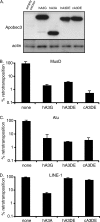
Fig. 1.
Fig. 1.
Human and chimpanzee Apobec3DE restrict retrotransposons. Human and chimpanzee Apobec3DE were expressed in retrotransposition assays to determine their antiviral activity. Retrotransposition activity is normalized to 100% in the absence of Apobec3 (none). (A) Western blot analysis of protein levels of human Apobec3G (hA3G), human Apobec3A (hA3A), human Apobec3DE (hA3DE), and chimpanzee Apobec3DE (cA3DE) during transient transfection. (B) Retrotransposition of MusD. Human Apobec3G was used as a positive control. (C) Retrotransposition of the prototypic Alu element. Human Apobec3A was used as a positive control. (D) Retrotransposition of LINE-1. Human Apobec3G was used as a positive control. Experiments were performed at least 3 times, and results from one representative experiment are shown. Error bars represent the standard deviation of triplicate transfections within one experiment.
Fig. 2.
Fig. 2.
Human and chimpanzee Apobec3DE restrict multiple Alu subfamilies. Human and chimpanzee Apobec3DE were expressed in retrotransposition assays to determine their antiviral activity. Retrotransposition activity is normalized to 100% in the absence of Apobec3 (none). (A) Retrotransposition of consensus Alu subfamilies in the absence or presence of human Apobec3G (hA3G), human Apobec3DE (hA3DE), or chimpanzee Apobec3DE (hA3DE). (B) Percentage of nonretroviral neomycin-resistant colonies formed in the absence or presence of human Apobec3DE. Error bars represent the standard deviation of triplicate transfections within one experiment.
Fig. 3.
Fig. 3.
Restriction of retroviruses by human and chimpanzee Apobec3DE. Human Apobec3DE (hA3DE) and chimpanzee Apobec3DE (cA3DE) were expressed in single-round infectivity assays to determine antiviral activity. Infections in the presence of human Apobec3G (hA3G) were used as a positive control. Infectivity is normalized to 100% in the absence of Apobec3 (none). (A) Infectivity of MLV. (B) Infectivity of HIV-2 without Vif (HIV-2Δvif) and of HIV-2 containing Vif (HIV-2 WT). (C) Infectivity of HIV-1 without Vif (HIVΔvif) and HIV-1 containing Vif (HIV WT). (D) Infectivity of SIVagmTAN without Vif (SIVagmTANΔvif) and SIVagmTAN containing Vif (SIVagmTAN WT). Experiments were performed at least 2 times, and results from one representative experiment are shown. Error bars represent the standard deviation of triplicate infections within one experiment. P values were calculated using paired two-tailed Student's t test.
Fig. 4.
Fig. 4.
Human and chimpanzee Apobec3DE are incorporated into HIV-1Δvif virions. The infectivity of HIV with and without Vif was assessed in the presence of increasing amounts of Apobec3DE. (A) Threefold dilutions of human and chimpanzee Apobec3DE were expressed in single-round infectivity assays. Infections are represented as a percentage of the infectivity of HIV-1 without Apobec3, which was set to 100%. Filled circles show infections in the presence of human Apobec3DE (hA3DE), and open circles show infections in the presence of chimpanzee Apobec3DE (cA3DE). Solid lines show infections with HIV-1 without Vif (HIV-1Δvif), and dashed lines show infections with HIV-1 containing Vif (HIV-1 WT). The experiment was performed at least 3 times, and results from one representative experiment are shown. Error bars represent the standard deviation of triplicate infections within one experiment. (B) Western blot analysis of Apobec3DE protein levels in the absence of Vif. (Top) Cell lysates from HIV-1Δvif infections. (Bottom) Viral lysates from HIV-1Δvif infections. (C) Western blot analysis of Apobec3DE protein levels in the presence of Vif. (Top) Cell lysates from infections with HIV-1 containing Vif. (Bottom) Viral lysates from infections with HIV-1 containing Vif. Apobec3DE levels are increasing from left to right on each blot. Apobec3DE was detected with an anti-HA antibody. Actin was used as a loading control for cellular lysates. Virions were normalized by quantification of p24 gag levels before lysis. (D) Pulse-chase analysis of Apobec3DE proteins. Human and chimpanzee Apobec3DE were radiolabeled for 30 min and chased for 0 min, 30 min, 1 h, and 20 h. Proteins were immunoprecipitated using an anti-HA antibody and resolved by SDS-PAGE. Autoradiography was used to visualize the gel.
Fig. 5.
Fig. 5.
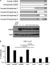
C terminus of Apobec3DE determines its ability to restrict HIV-1Δvif. (A) (Top) Schematic of human/chimpanzee (h/c) Apobec3DE chimeras used in infectivity assays. (Bottom) Western blot analysis of cellular levels of Apobec3DE chimeras. Apobec3DE was detected with an anti-HA antibody. Actin was used as a loading control. (B) Infectivity of HIV-1Δvif in the presence of human/chimpanzee chimeras. Infections are represented as a percentage of the infectivity of HIV-1 without Apobec3 (none), which was set to 100%. Experiments were performed at least 3 times, and results from one representative experiment are shown. Error bars represent the standard deviation of triplicate infections within one experiment. P values were calculated using two-tailed Student's t test.
Fig. 6.
Fig. 6.
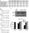
Chimpanzee Apobec3DE induces higher levels of hypermutation than human Apobec3DE does during viral infection. (A) HIV-1 genomes were sequenced from HIV-1Δvif infections performed in the absence of Apobec3 (none) or in the presence of human Apobec3G (hA3G), human Apobec3DE (hA3DE), chimpanzee Apobec3DE (cA3DE), or human/chimpanzee chimeras, and the G-to-A mutation rate of viral genomes was calculated. The rate of other mutations was also calculated. Combined data from three independent experiments are shown. (B) The nucleotide context of G-to-A mutations was characterized. The percentage of each nucleotide occurring at the −2, −1, +1, and +2 positions of deaminated cytidines is shown, with the most common nucleotide listed below each position. The number of mutations examined is noted in the top left corner of each grid. (C) (Top) Western blot analysis of cellular levels of human (h) and chimpanzee (c) Apobec3DE catalytic mutants. Apobec3DE was detected with an anti-HA antibody. Actin was used as a loading control. (Bottom) Infectivity of HIV-1Δvif in the presence of wild-type Apobec3DE (WT) or Apobec3DE catalytic mutants (E264Q and E80Q). Infections are represented as a percentage of the infectivity of HIV-1 without Apobec3 (none), which was set to 100%. Experiments were performed at least 2 times, and results from one representative experiment are shown. Error bars represent the standard deviation of triplicate infections within one experiment.
Fig. 7.
Fig. 7.
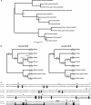
Apobec3DE evolved rapidly in chimpanzee ancestors. Sequences obtained by PCR in this study, as described in Materials and Methods, were analyzed. (A) A maximum-likelihood phylogeny based on primate Apobec3DE and Apobec3F sequences is shown. Statistical support, calculated as aLRT values, is shown for each node. (B) Positive selection analysis of Apobec3DE. Two phylogenies of Apobec3DE, one with the human ortholog as an outgroup to gorilla, chimpanzee, and bonobo Apobec3DE (right) and another with gorilla Apobec3DE as an outgroup to human, chimpanzee, and bonobo Apobec3DE (left), were analyzed. Global dN/dS ratios are indicated above each branch. The numbers of nonsynonymous and synonymous changes along each branch are indicated below each branch in parentheses (NS:S). dN/dS values greater than 1 are indicative of positive selection. (C) Codons in Apobec3DE under positive selection. An alignment of human and chimpanzee Apobec3DE protein sequences is shown with chimpanzee-specific changes highlighted in black. Residues having a high probability of being under positive selection across all primates are marked with asterisks. Residues with BEB values of >0.95 are marked with a single asterisk, and residues with BEB values of >0.99 are marked with two asterisks. The C-terminal domain in Apobec3DE that was tested for antiviral activity is underlined, with the minimal 5 residues that are required for antiviral activity underlined twice.

Similar articles

Cited by

References

    1. Aguiar R. S., Lovsin N., Tanuri A., Peterlin B. M. 2008. Vpr.A3A chimera inhibits HIV replication. J. Biol. Chem. 283:2518–2525 - PubMed
    1. Alisch R. S., Garcia-Perez J. L., Muotri A. R., Gage F. H., Moran J. V. 2006. Unconventional translation of mammalian LINE-1 retrotransposons. Genes Dev. 20:210–224 - PMC - PubMed
    1. Bartz S. R., Vodicka M. A. 1997. Production of high-titer human immunodeficiency virus type 1 pseudotyped with vesicular stomatitis virus glycoprotein. Methods 12:337–342 - PubMed
    1. Batzer M. A., Deininger P. L. 2002. Alu repeats and human genomic diversity. Nat. Rev. Genet. 3:370–379 - PubMed
    1. Bennett E. A., et al. 2008. Active Alu retrotransposons in the human genome. Genome Res. 18:1875–1883 - PMC - PubMed

Publication types

LinkOut - more resources