. 2011 Oct;85(20):10649-58.
doi: 10.1128/JVI.05034-11. Epub 2011 Aug 10.

Comparisons between murine polyomavirus and Simian virus 40 show significant differences in small T antigen function

Affiliations

Comparisons between murine polyomavirus and Simian virus 40 show significant differences in small T antigen function

Shaida Andrabi et al. J Virol. 2011 Oct.

Abstract

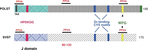
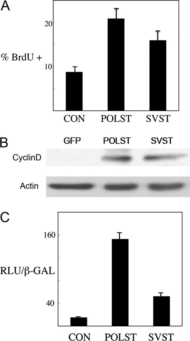
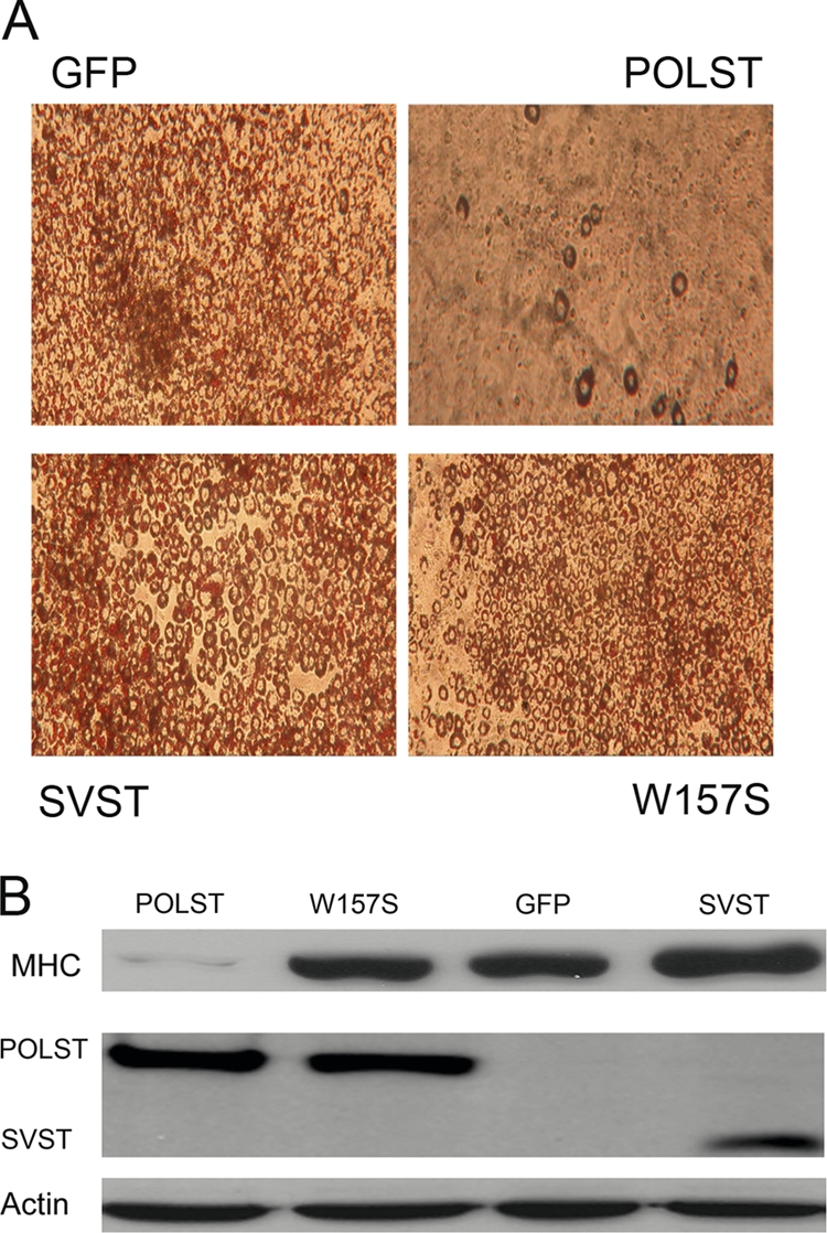
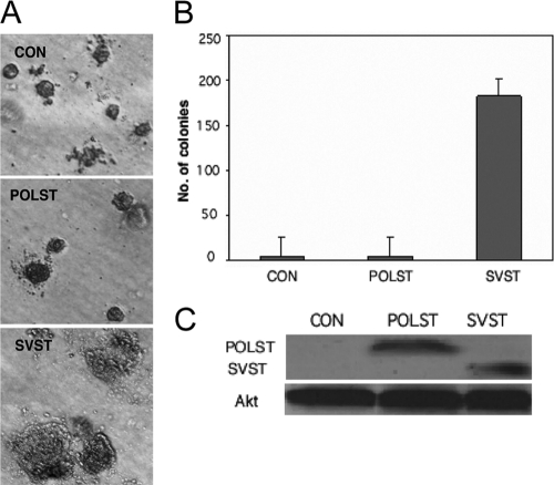
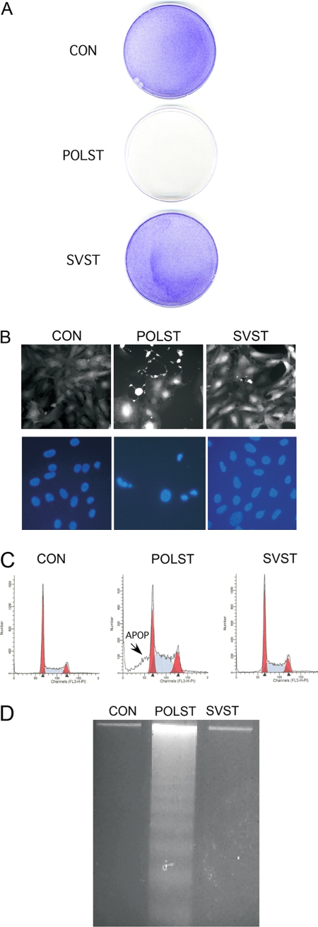
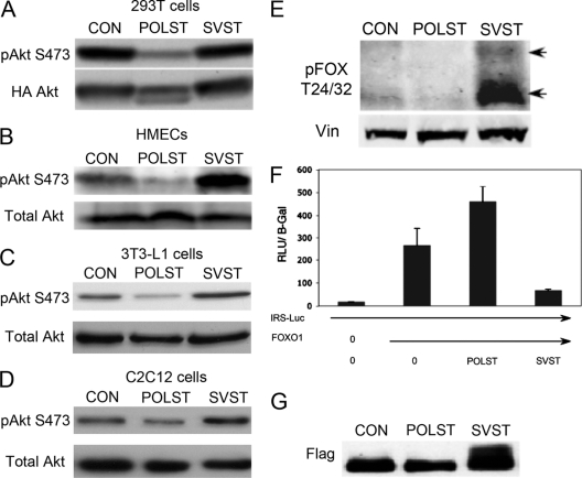
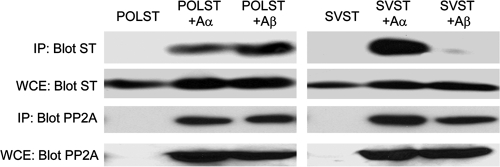
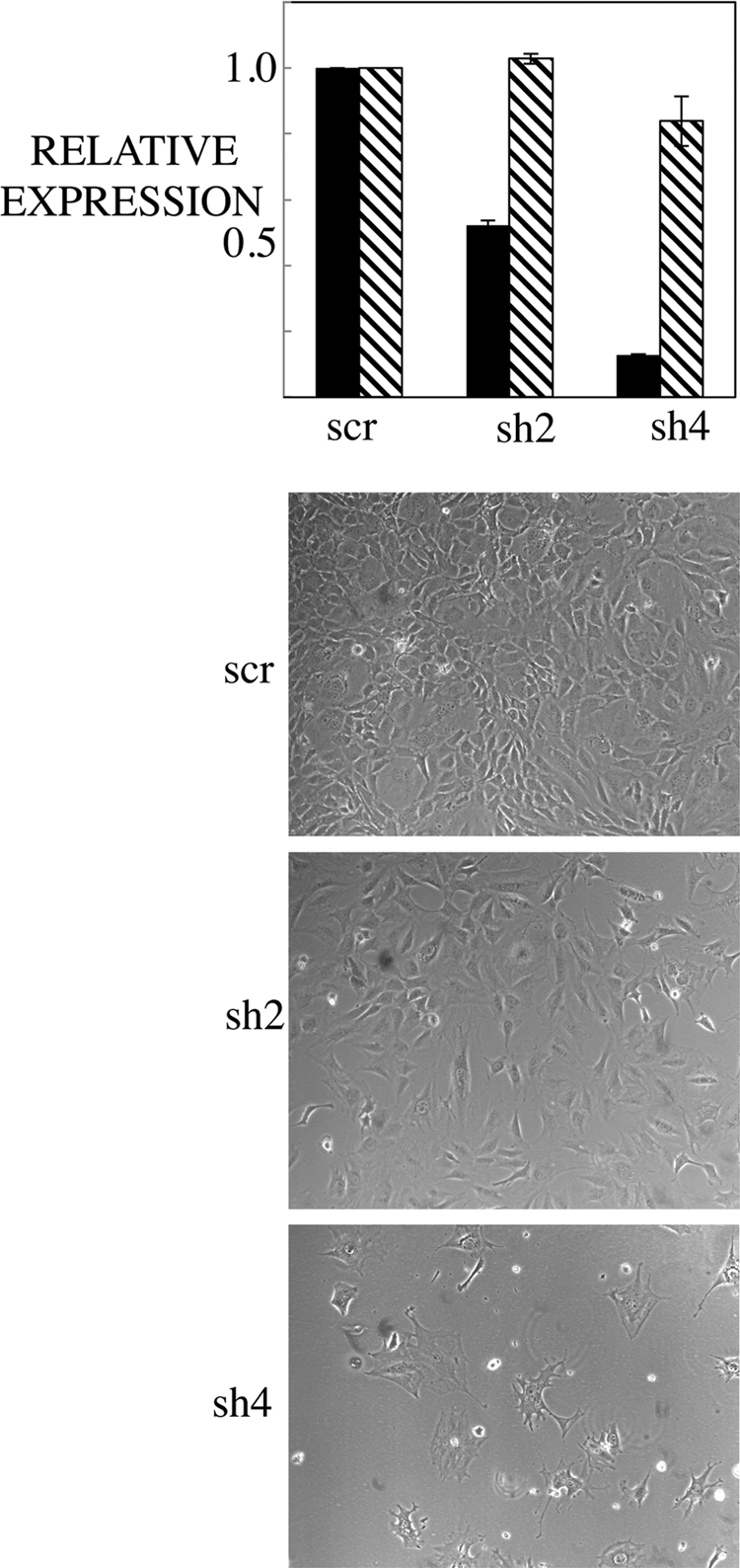
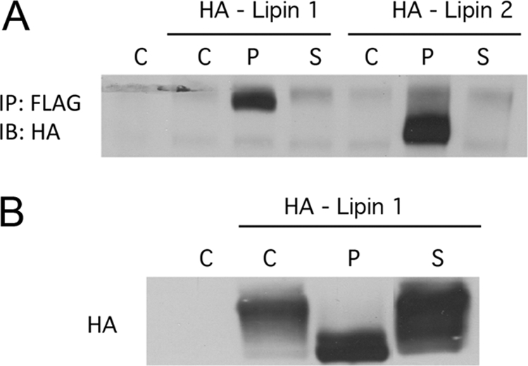
Although members of a virus family produce similar gene products, those products may have quite different functions. Simian virus 40 (SV40) large T antigen (LT), for example, targets p53 directly, but murine polyomavirus LT does not. SV40 small T antigen (SVST) has received considerable attention because of its ability to contribute to transformation of human cells. Here, we show that there are major differences between SVST and polyomavirus small T antigen (POLST) in their effects on differentiation, transformation, and cell survival. Both SVST and POLST induce cell cycle progression. However, POLST also inhibits differentiation of 3T3-L1 preadipocytes and C2C12 myoblasts. Additionally, POLST induces apoptosis of mouse embryo fibroblasts. SVST reduces the proapoptotic transcriptional activity of FOXO1 through phosphorylation. On the other hand, SVST complements large T antigen and Ras for the transformation of human mammary epithelial cells (HMECs), but POLST does not. Mechanistically, the differences between SVST and POLST may lie in utilization of protein phosphatase 2A (PP2A). POLST binds both Aα and Aβ scaffolding subunits of PP2A while SVST binds only Aα. Knockdown of Aβ could mimic POLST-induced apoptosis. The two small T antigens can target different proteins for dephosphorylation. POLST binds and dephosphorylates substrates, such as lipins, that SVST does not.


Figures

Fig. 1.
Fig. 1.
Schematic representation of polyomavirus ST (POLST) and SV40 ST (SVST) protein structures with the main features indicated.
Fig. 2.
Fig. 2.
Cell cycle-related effects of STs. (A) Effect of STs on S-phase progression. NIH 3T3 cells expressing control vector (CON), POLST, or SVST, as obtained by retroviral infection, were grown overnight in medium containing 0.2% serum. Cells were then incubated in medium containing BrdU for 1 h and subjected to immunofluorescence. Percent cells undergoing S phase (% BrdU +) is measured by BrdU incorporation compared to the total number of cells, as measured by DAPI-stained nuclei. (B) After retroviral infection C2C12 myoblast cell pools were isolated that express GFP, POLST, or SVST. These respective cells were grown to around 60% confluence, and fresh medium containing 10% FCS was added to the cells at this point for 4 hours. Cell lysates were subsequently collected and analyzed by Western blotting for cyclin D expression using actin as a control. (C) Effect of STs on the cyclin D1 promoter. NIH 3T3 cells were cotransfected with cyclin D-luc reporter, small T antigens, or control vector and pCMV-β-Gal. The luciferase activity was normalized using β-galactosidase activity, and results are reported as the number of relative light units (RLU) × 10−3/β-galactosidase activity unit.
Fig. 3.
Fig. 3.
POLST inhibits differentiation of 3T3-L1 preadipocytes and C2C12 myoblasts. (A) After retroviral infection 3T3-L1 cell pools were isolated that expressed GFP, POLST, W157S POLST, or SVST. Subsequently, cells were allowed to differentiate. At 7 days postinduction, differentiating cells were fixed and stained with Oil Red O. (B) After retroviral infection C2C12 myoblast cell pools were isolated that expressed GFP, POLST, PP2A W157S POLST, or SVST. Cell lysates were collected at 6 days postinduction of differentiation. Expression of MHC, a differentiation marker, was determined by Western blotting. PN116 antibody was used to detect the expression of POLST, while PAB419 antibody was used to detect the expression of SVST. α-Actin was used as a loading control.
Fig. 4.
Fig. 4.
Effect of STs on transformation measured by anchorage-independent growth. (A) HMECs that express hTERT, SV40 large T antigen, and H-ras were used as controls (CON) in soft-agar assays. POLST or SVST cells were then expressed. Colonies are shown after 6 weeks of growth. (B) Quantitative representation of the soft-agar assay. Colonies 0.2 mm or more in size were counted after 6 weeks of growth. (C) Cell extracts from HMECs were separated by PAGE and blotted with antibodies against POLST (PN116) or SVST (PAB419) to determine the relative expression levels of the STs. Total Akt was used as a loading control.
Fig. 5.
Fig. 5.
Effect of STs on cell survival. NIH 3T3 cells expressing small T antigens were obtained by retroviral infection. (A) Cell survival following retroviral infection. Cells were infected with retrovirus containing only the puromycin resistance gene (pBABE-puro) as a control (CON) expressing POLST (pBABEpuro-POLST) or SVST (pBABEpuro-SVST). After selection in puromycin (2.5 μg/ml) for about 7 days, cells were fixed with ethanol, stained with 0.2% crystal violet, and washed (78). (B) Expression of STs was confirmed by GFP expression as seen in fluorescence microscopy, and the morphology of the cells expressing POLST indicated cell death. Nuclei were stained by DAPI and observed by fluorescence microscopy. (C) POLST induced apoptosis (APOP) as seen by FACS analysis. A sub-G1 peak was seen in the cell cycle histogram of POLST-expressing cells but not in control (CON) or SVST-expressing cells. FACS analysis showed that 37% of the infected cells with POLST had sub-G1 DNA content. (D) Apoptosis in POLST-expressing cells as indicated by DNA fragmentation. DNA was extracted from ST-expressing NIH 3T3 cells, separated on a 2% agarose gel, stained with ethidium bromide, and visualized on a UV transilluminator.
Fig. 6.
Fig. 6.
ST effects on Akt signaling. (A) Effects of ST on Akt S473 phosphorylation. 293T cells were transiently cotransfected with HA-myr-Akt (HA-labeled myristoylated Akt) and pCMV-ST antigens. At 48 h posttransfection, cell extracts were blotted for phospho-Akt (S473) or HA (total Akt) as indicated. (B to D) Effects of overexpression of STs on endogenous Akt S473 phosphorylation. Extracts from control HMECs, 3T3-L1s, and C2C12s cells (CON) and those expressing either POLST or SVST were collected. Phosphorylation of endogenous Akt at S473 was detected by Western blotting. Total Akt was used as a loading control in each case. (E) Effects of overexpression of STs on FOXO1/FOXO3a phosphorylation. Extracts from control HMECs (CON) and those expressing either POLST or SVST were collected for Western blotting. Phospho-FOXO1/FOXO3a (T24/32) was detected with vinculin as a loading control. (F) Effects of STs on FOXO1 activation. 293T cells were transiently transfected with IRS-Luc, Flag-FOXO1, pCMV–β-Gal and either POLST or SVST. Extracts were obtained after 24 h and used for luciferase and β-Gal assays. The luciferase values (RLU × 10−3) were normalized by β-galactosidase values. Standard errors of the means are shown. (G) Flag-FOXO1 mobility was determined using anti-FLAG antibody after cotransfection with control vector, POLST, or SVST as described for panel F.
Fig. 7.
Fig. 7.
Interaction of small T antigens with PP2A A isoforms. NIH 3T3 cells were cotransfected with EE-tagged PP2A Aα or Aβ and POLST or SVST. At 48 h after transfection, lysates were collected, and PP2A Aα or Aβ was immunoprecipitated (IP) with anti-EE antibody and blotted for small T antigens. Expression of small T antigens and PP2A was confirmed by Western blotting on whole-cell extracts (WCE).
Fig. 8.
Fig. 8.
Short hairpin knockdown of PP2A Aβ induces cell death. 3T3-L1 cells were infected with pLKO control (scr, for scrambled) or sh2 Aβ or sh4 Aβ. At 48 h postinfection, mRNA was collected from the cells. RT-PCR was performed to assay for levels of Aβ (black bars) and the puromycin resistance gene (PAC gene; striped bars). Phase-contrast pictures were taken at 7 days postinfection (magnification, ×10).
Fig. 9.
Fig. 9.
Interaction of STs with lipins. (A) Interaction of ST with both lipin 1 and lipin 2. 293T cells were cotransfected with either HA-lipin 1 or lipin 2 and empty vector (C), FLAG-POLST (P), or FLAG-SVST (S). STs were immunoprecipitated (IP) with anti-FLAG antibody, and the immunoprecipitates were blotted for lipins with anti-HA (IB). (B) The effect of STs on lipin 1 mobility. 293T cells were cotransfected with HA-lipin 1 and vector (C), POLST (P), or SVST (S). After approximately 40 h, cells extracts were collected and blotted for lipin using anti-HA antibody.

Similar articles

Cited by

References

    1. Altiok S., Xu M., Spiegelman B. M. 1997. PPARγ induces cell cycle withdrawal: inhibition of E2F/DP DNA-binding activity via down-regulation of PP2A. Genes Dev. 11:1987–1998 - PMC - PubMed
    1. Andrabi S., Gjoerup O. V., Kean J. A., Roberts T. M., Schaffhausen B. 2007. Protein phosphatase 2A regulates life and death decisions via Akt in a context-dependent manner. Proc. Natl. Acad. Sci. U. S. A. 104:19011–19016 - PMC - PubMed
    1. Asselin C., Gelinas C., Bastin M. 1983. Role of the three polyoma virus early proteins in tumorigenesis. Mol. Cell. Biol. 3:1451–1459 - PMC - PubMed
    1. Asselin C., Vass-Marengo J., Bastin M. 1986. Mutation in the polyomavirus genome that activates the properties of large T associated with neoplastic transformation. J. Virol. 57:165–172 - PMC - PubMed
    1. Berger H., Wintersberger E. 1986. Polyomavirus small T antigen enhances replication of viral genomes in 3T6 mouse fibroblasts. J. Virol. 60:768–770 - PMC - PubMed

Publication types

Substances