

Exosomes from human CD34(+) stem cells mediate their proangiogenic paracrine activity
- PMID: 21835908
- PMCID: PMC3201702
- DOI: 10.1161/CIRCRESAHA.111.253286
Exosomes from human CD34(+) stem cells mediate their proangiogenic paracrine activity
Abstract
Rationale: Transplantation of human CD34(+) stem cells to ischemic tissues has been associated with reduced angina, improved exercise time, and reduced amputation rates in phase 2 clinical trials and has been shown to induce neovascularization in preclinical models. Previous studies have suggested that paracrine factors secreted by these proangiogenic cells are responsible, at least in part, for the angiogenic effects induced by CD34(+) cell transplantation.
Objective: Our objective was to investigate the mechanism of CD34(+) stem cell-induced proangiogenic paracrine effects and to examine if exosomes, a component of paracrine secretion, are involved.
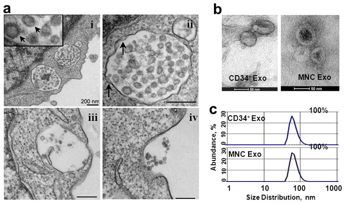
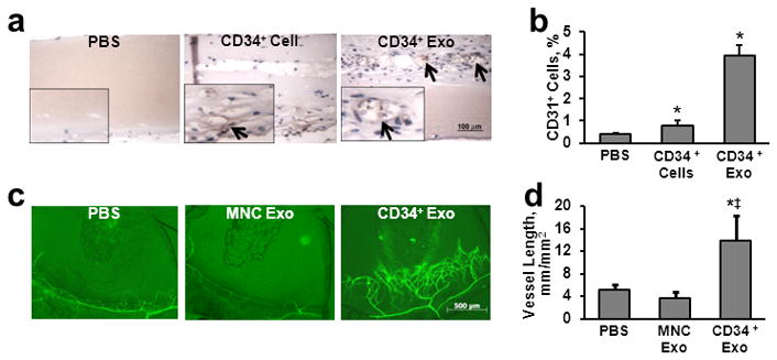
Methods and results: Exosomes collected from the conditioned media of mobilized human CD34(+) cells had the characteristic size (40 to 90 nm; determined by dynamic light scattering), cup-shaped morphology (electron microscopy), expressed exosome-marker proteins CD63, phosphatidylserine (flow cytometry) and TSG101 (immunoblotting), besides expressing CD34(+) cell lineage marker protein, CD34. In vitro, CD34(+) exosomes replicated the angiogenic activity of CD34(+) cells by increasing endothelial cell viability, proliferation, and tube formation on Matrigel. In vivo, the CD34(+) exosomes stimulated angiogenesis in Matrigel plug and corneal assays. Interestingly, exosomes from CD34(+) cells but not from CD34(+) cell-depleted mononuclear cells had angiogenic activity.
Conclusions: Our data demonstrate that human CD34(+) cells secrete exosomes that have independent angiogenic activity both in vitro and in vivo. CD34(+) exosomes may represent a significant component of the paracrine effect of progenitor cell transplantation for therapeutic angiogenesis.
Figures


References
-
- Losordo D. Randomized double-blind, placebo controlled trial of autologous cd34+ cell therapy for critical limb ischemia: 1 year results. Circulation. 2010;122:A16920.
-
- Kawamoto A, Iwasaki H, Kusano K, Murayama T, Oyamada A, Silver M, Hulbert C, Gavin M, Hanley A, Ma H, Kearney M, Zak V, Asahara T, Losordo DW. Cd34-positive cells exhibit increased potency and safety for therapeutic neovascularization after myocardial infarction compared with total mononuclear cells. Circulation. 2006;114:2163–2169. - PubMed
-
- Asahara T, Murohara T, Sullivan A, Silver M, van der Zee R, Li T, Witzenbichler B, Schatteman G, Isner JM. Isolation of putative progenitor endothelial cells for angiogenesis. Science. 1997;275:964–967. - PubMed
-
- Kumar AH, Caplice NM. Clinical potential of adult vascular progenitor cells. Arterioscler Thromb Vasc Biol. 2010;30:1080–1087. - PubMed
Publication types
MeSH terms
Substances
Grants and funding
- R01 HL077428/HL/NHLBI NIH HHS/United States
- R01 HL080137/HL/NHLBI NIH HHS/United States
- R01 HL057516/HL/NHLBI NIH HHS/United States
- P01 HL108795/HL/NHLBI NIH HHS/United States
- 1P01HL108795/HL/NHLBI NIH HHS/United States
- 5R01HL077428/HL/NHLBI NIH HHS/United States
- R01 HL095874/HL/NHLBI NIH HHS/United States
- R01 AR054796/AR/NIAMS NIH HHS/United States
- 5R01HL095874/HL/NHLBI NIH HHS/United States
- R01 HL053354/HL/NHLBI NIH HHS/United States
- 2R01HL053354/HL/NHLBI NIH HHS/United States
- R01 AR050250/AR/NIAMS NIH HHS/United States
LinkOut - more resources
Full Text Sources
Other Literature Sources
Medical
Molecular Biology Databases
Miscellaneous
