

Differentiation of brain abscesses from necrotic glioblastomas and cystic metastatic brain tumors with diffusion tensor imaging
- PMID: 21835939
- PMCID: PMC7965370
- DOI: 10.3174/ajnr.A2581
Differentiation of brain abscesses from necrotic glioblastomas and cystic metastatic brain tumors with diffusion tensor imaging
Abstract
Background and purpose: The differentiation of abscesses from glioblastomas and metastases may not always be possible on the basis of DWI. Our hypothesis was that differences in diffusion properties as detected by DTI allow differentiation of abscess from glioblastomas and metastasis. Furthermore, diagnostic performance of tensor metrics quantifying anisotropy or tensor shapes is better than that of ADC in measuring mean diffusivity for this purpose.
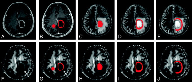
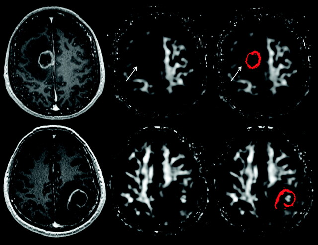
Materials and methods: DTI was performed in 15 abscesses, 15 necrotic glioblastomas, and 26 cystic metastases. In each lesion, manually segmented into 4 regions of interest (ie, cystic cavity, enhancing rim, and immediate [edema most adjacent to the enhancing rim] and distant zones of edema), FA, ADC, C(l), C(p), and C(s) values were measured and statistically compared among groups and evaluated with ROC curve analysis. The presence of a hyperintense FA rim (a rim of edematous tissue that was hyperintense on the FA map) was assessed visually.
Results: Abscess was significantly different from glioblastoma for all tensor metrics measured in the cystic cavity and immediate zone of edema and for all except C(l) in the enhancing rim. Abscess was significantly different from metastasis for all tensor metrics measured in the cystic cavity and enhancing rim and for FA, ADC, and C(l) in immediate zone of edema. The incidence of a hyperintense FA rim was significantly higher in glioblastoma and metastasis compared with abscess. The 3 tensor metrics with the highest performance in differentiating abscess from glioblastoma and metastasis were FA, C(l), and C(s) of the cystic cavity.
Conclusions: DTI is able to differentiate abscess from glioblastoma and metastasis. FA, C(l), and C(s) outperformed ADC in diagnostic performance comparisons.
Figures
Similar articles
-
Diagnostic performance of regional DTI-derived tensor metrics in glioblastoma multiforme: simultaneous evaluation of p, q, L, Cl, Cp, Cs, RA, RD, AD, mean diffusivity and fractional anisotropy.Eur Radiol. 2013 Apr;23(4):1112-21. doi: 10.1007/s00330-012-2688-7. Epub 2012 Oct 21. Eur Radiol. 2013. PMID: 23085868
-
Differential diagnosis of intracranial ring enhancing cystic mass lesions--role of diffusion-weighted imaging (DWI) and diffusion-tensor imaging (DTI).Clin Neurol Neurosurg. 2010 Apr;112(3):218-25. doi: 10.1016/j.clineuro.2009.11.016. Epub 2010 Jan 6. Clin Neurol Neurosurg. 2010. PMID: 20053496
-
Susceptibility-weighted imaging provides complementary value to diffusion-weighted imaging in the differentiation between pyogenic brain abscesses, necrotic glioblastomas, and necrotic metastatic brain tumors.Eur J Radiol. 2019 Aug;117:56-61. doi: 10.1016/j.ejrad.2019.05.021. Epub 2019 May 30. Eur J Radiol. 2019. PMID: 31307653
-
The role of diffusion tensor imaging and fractional anisotropy in the evaluation of patients with idiopathic normal pressure hydrocephalus: a literature review.Neurosurg Focus. 2016 Sep;41(3):E12. doi: 10.3171/2016.6.FOCUS16192. Neurosurg Focus. 2016. PMID: 27581308 Review.
-
Diffusion-weighted MR imaging in the preoperative assessment of brain abscesses.Surg Neurol. 2002 Dec;58(6):395-402; discussion 402. doi: 10.1016/s0090-3019(02)00929-1. Surg Neurol. 2002. PMID: 12517619 Review.
Cited by
-
Advances in the application of neuroinflammatory molecular imaging in brain malignancies.Front Immunol. 2023 Jul 18;14:1211900. doi: 10.3389/fimmu.2023.1211900. eCollection 2023. Front Immunol. 2023. PMID: 37533851 Free PMC article. Review.
-
Diagnostic utility of diffusion tensor imaging in differentiating glioblastomas from brain metastases.AJNR Am J Neuroradiol. 2014 May;35(5):928-34. doi: 10.3174/ajnr.A3871. Epub 2014 Feb 6. AJNR Am J Neuroradiol. 2014. PMID: 24503556 Free PMC article.
-
Diagnostic performance of regional DTI-derived tensor metrics in glioblastoma multiforme: simultaneous evaluation of p, q, L, Cl, Cp, Cs, RA, RD, AD, mean diffusivity and fractional anisotropy.Eur Radiol. 2013 Apr;23(4):1112-21. doi: 10.1007/s00330-012-2688-7. Epub 2012 Oct 21. Eur Radiol. 2013. PMID: 23085868
-
Classification of brain lesions using a machine learning approach with cross-sectional ADC value dynamics.Sci Rep. 2023 Jul 15;13(1):11459. doi: 10.1038/s41598-023-38542-7. Sci Rep. 2023. PMID: 37454179 Free PMC article.
-
Clinical applications of diffusion-weighted sequence in brain imaging: beyond stroke.Neuroradiology. 2022 Jan;64(1):15-30. doi: 10.1007/s00234-021-02819-3. Epub 2021 Oct 1. Neuroradiology. 2022. PMID: 34596716 Free PMC article. Review.
References
-
- Reddy JS, Mishra AM, Behari S, et al. . The role of diffusion-weighted imaging in the differential diagnosis of intracranial cystic mass lesions: a report of 147 lesions. Surg Neurol 2006; 66: 246–50 - PubMed
-
- Lee EJ, Ahn KJ, Ha YS, et al. . Unusual findings in cerebral abscess: report of two cases. Br J Radiol 2006; 79: e156–61 - PubMed
-
- Mishra AM, Gupta RK, Jaggi RS, et al. . Role of diffusion-weighted imaging and in vivo proton magnetic resonance spectroscopy in the differential diagnosis of ring-enhancing intracranial cystic mass lesions. J Comput Assist Tomogr 2004; 28: 540–47 - PubMed
-
- Holtas S, Geijer B, Stromblad LG, et al. . A ring-enhancing metastasis with central high signal on diffusion-weighted imaging and low apparent diffusion coefficients. Neuroradiology 2000; 42: 824–27 - PubMed
Publication types
MeSH terms
LinkOut - more resources
Full Text Sources
Medical
Miscellaneous