

Mild cognitive impairment: differential atrophy in the hippocampal subfields
- PMID: 21835940
- PMCID: PMC3268157
- DOI: 10.3174/ajnr.A2589
Mild cognitive impairment: differential atrophy in the hippocampal subfields
Abstract
Background and purpose: Hippocampus volumetry is a useful surrogate marker for the diagnosis of Alzheimer disease, but it seems insufficiently sensitive for the aMCI stage. We postulated that some hippocampus subfields are specifically atrophic in aMCI and that measuring hippocampus subfield volumes will improve sensitivity of MR imaging to detect aMCI.
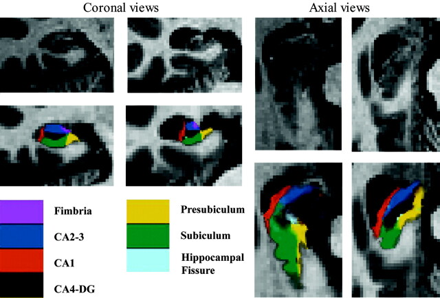
Materials and methods: We evaluated episodic memory and hippocampus subfield volume in 15 patients with aMCI and 15 matched controls. After segmentation of the whole hippocampus from clinical MR imaging, we applied a new computational method allowing fully automated segmentation of the hippocampus subfields. This method used a Bayesian modeling approach to infer segmentations from the imaging data.
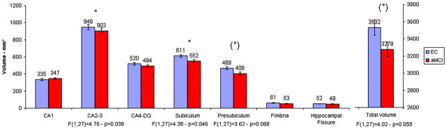
Results: In comparison with controls, subiculum and CA2-3 were significantly atrophic in patients with aMCI, whereas total hippocampus volume and other subfields were not. Total hippocampus volume in controls was age-related, whereas episodic memory was the main explanatory variable for both the total hippocampus volume and the subfields that were atrophic in patients with aMCI. Segmenting subfields increases sensitivity to diagnose aMCI from 40% to 73%.
Conclusions: Measuring CA2-3 and subiculum volumes allows a better detection of aMCI.
Figures
References
-
- Petersen RC. Mild cognitive impairment as a diagnostic entity. J Intern Med 2004; 256: 183–94 - PubMed
-
- Dubois B, Feldman HH, Jacova C, et al. Research criteria for the diagnosis of Alzheimer's disease: revising the NINCDS-ADRDA criteria. Lancet Neurol 2007; 6: 734–46 - PubMed
-
- Giannakopoulos P, Kovari E, Gold G, et al. Pathological substrates of cognitive decline in Alzheimer's disease. Front Neurol Neurosci 2009; 24: 20–29 - PubMed
-
- Frisoni GB, Ganzola R, Canu E, et al. Mapping local hippocampal changes in Alzheimer's disease and normal ageing with MRI at 3 Tesla. Brain 2008; 131: 3266–76 - PubMed
Publication types
MeSH terms
Grants and funding
LinkOut - more resources
Full Text Sources
Medical
Miscellaneous