. 2011 Aug 12;333(6044):883-5.
doi: 10.1126/science.1204824.

The structure of the kinesin-1 motor-tail complex reveals the mechanism of autoinhibition

Affiliations

The structure of the kinesin-1 motor-tail complex reveals the mechanism of autoinhibition

Hung Yi Kristal Kaan et al. Science. .

Abstract

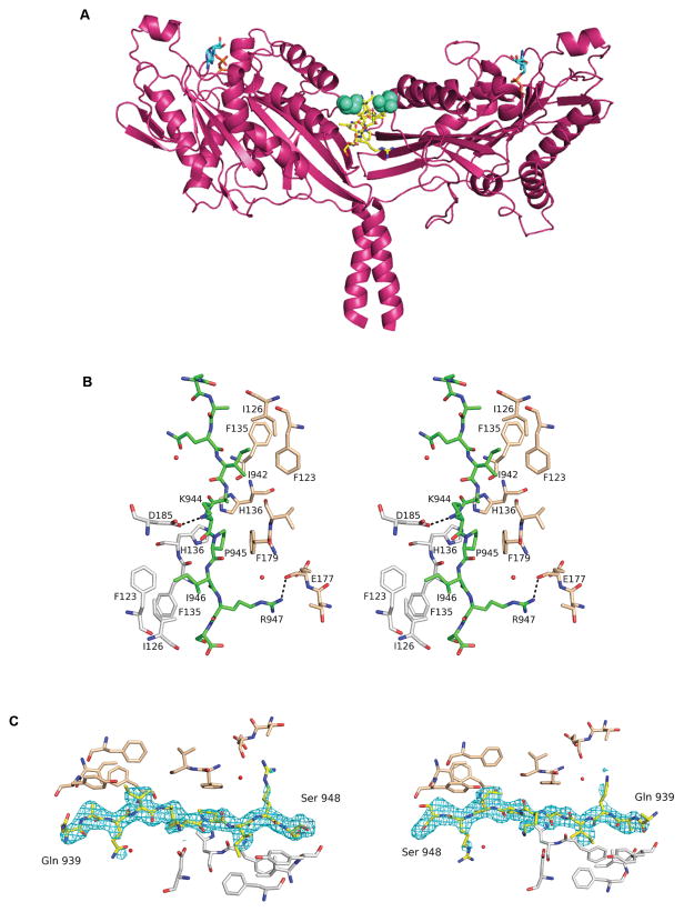
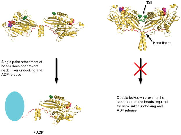
When not transporting cargo, kinesin-1 is autoinhibited by binding of a tail region to the motor domains, but the mechanism of inhibition is unclear. We report the crystal structure of a motor domain dimer in complex with its tail domain at 2.2 angstroms and compare it with a structure of the motor domain alone at 2.7 angstroms. These structures indicate that neither an induced conformational change nor steric blocking is the cause of inhibition. Instead, the tail cross-links the motor domains at a second position, in addition to the coiled coil. This "double lockdown," by cross-linking at two positions, prevents the movement of the motor domains that is needed to undock the neck linker and release adenosine diphosphate. This autoinhibition mechanism could extend to some other kinesins.


Figures

Figure 1
Figure 1. Structure of motor-tail complex
(A) D. melanogaster kinesin motor domain dimer complexed with tail domain (yellow). (Cyan sticks: Mg2+ADP, green spheres: Ser 181). (B) Stereoplot showing C—H-π and hydrogen bond interactions (dotted lines) between the tail (green) and motor domains (chain A: grey, B: beige). (C) Dimer-tail interface showing residues of motor domain (chain A: grey, B: beige), tail domain (yellow) and omit map (Fo-Fc) (contoured at σ=3.00) for tail binding in both directions.
Figure 2
Figure 2. Disulfide mimic in mant-ADP release experiment
Cys-lite motor domain dimer with a S181C substitution was oxidized to the covalent disulfide dimer and then reduced back to the sulfhydryl form by addition of dithiothreitol (DTT). (A) Time course for release of mant-ADP from unmodified (SH), oxidized (SS), and DTT treated (SS + DTT) species after mixing with a chase of excess unlabeled ATP. Concentrations after mixing were 0.15 3M kinesin motor domain (0.075 3M dimer) with or without 0.3 3M tubulin (dimer) as indicated. (B) Formation of the covalently linked disulphide dimer was analyzed by denaturing SDS-PAGE in the absence of reducing agents.
Figure 3
Figure 3. Proposed model for ‘double lockdown’ mechanism of autoinhibition
Based on the crystal structures, the motor domains (yellow, cartoon) have considerable freedom of movement for neck linker (red) undocking and ADP (red, spheres) release in the absence of tail (green, sticks) binding. Cyan oval represents nucleotide-free motor domain with neck-linker undocked. However, when the tail binds, the motor domains are also cross-linked at the tail interface. In light of the cross-linking experiment results and based on the neck linker undocking hypothesis, we propose a ‘double lockdown’ mechanism, which prevents the separation of the motor domains that is required for neck linker undocking and ADP release. Ser 181 residues (green, spheres) are far apart in the free dimer but close together in the dimer-tail complex, and are on opposite sides of the tail interface.

Similar articles

Cited by

References

    1. Vale RD, Reese TS, Sheetz MP. Cell. 1985 Aug;42:39. - PMC - PubMed
    1. Hirokawa N. Science. 1998 Jan 23;279:519. - PubMed
    1. Endow SA, Kull FJ, Liu H. J Cell Sci. 2010 Oct 15;123:3420. - PMC - PubMed
    1. Hackney DD, Stock MF. Nat Cell Biol. 2000 May;2:257. - PubMed
    1. Stock MF, et al. J Biol Chem. 1999 May 21;274:14617. - PubMed

Publication types