. 2011 Aug 23;108(34):14121-6.
doi: 10.1073/pnas.1103621108. Epub 2011 Aug 11.

Macromolecular organization of ATP synthase and complex I in whole mitochondria

Affiliations

Macromolecular organization of ATP synthase and complex I in whole mitochondria

Karen M Davies et al. Proc Natl Acad Sci U S A. .

Abstract

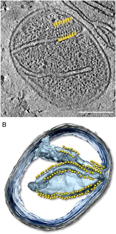
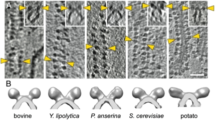
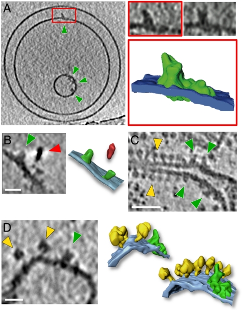
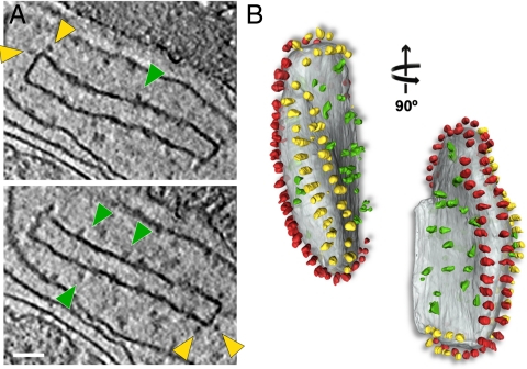
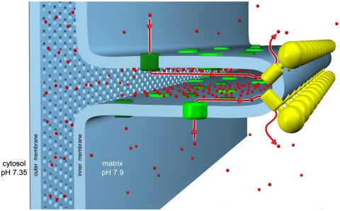
We used electron cryotomography to study the molecular arrangement of large respiratory chain complexes in mitochondria from bovine heart, potato, and three types of fungi. Long rows of ATP synthase dimers were observed in intact mitochondria and cristae membrane fragments of all species that were examined. The dimer rows were found exclusively on tightly curved cristae edges. The distance between dimers along the rows varied, but within the dimer the distance between F(1) heads was constant. The angle between monomers in the dimer was 70° or above. Complex I appeared as L-shaped densities in tomograms of reconstituted proteoliposomes. Similar densities were observed in flat membrane regions of mitochondrial membranes from all species except Saccharomyces cerevisiae and identified as complex I by quantum-dot labeling. The arrangement of respiratory chain proton pumps on flat cristae membranes and ATP synthase dimer rows along cristae edges was conserved in all species investigated. We propose that the supramolecular organization of respiratory chain complexes as proton sources and ATP synthase rows as proton sinks in the mitochondrial cristae ensures optimal conditions for efficient ATP synthesis.


Conflict of interest statement

The authors declare no conflict of interest.

Figures

Fig. 1.
Fig. 1.
Rows of F1-Fo ATP synthase dimers in whole mitochondria. (A) Tomographic slice through a whole mitochondrion of Podospora anserina showing arrays of dimeric ATP synthases (yellow arrowheads). (B) Segmented surface representation of A showing position of ATP synthase dimers (yellow spheres) to cristae membrane (blue). Dimers are confined to the highly curved cristae edges (see Movie S1). The mitochondrion is intact, as shown by the granular appearance of the matrix, in which ribosomes are visible as dark gray, approximately 25-nm particles. Gray, outer membrane; blue, cristae membranes; blue-transparent, inner boundary membrane; yellow, ATP synthase. Scale bar, 200 nm.
Fig. 2.
Fig. 2.
Rows of F1-Fo ATP synthase dimers from five different species. (A) Tomographic slices showing linear arrays of F1-Fo ATP synthase dimers in mitochondrial membranes from bovine heart, Yarrowia lipolytica, Podospora anserina, Saccharomyces cerevisiae, and potato. (Inset) Side view of each array showing dimers in relation to the membrane. Yellow arrowheads indicate F1 heads of one dimer. Scale bar, 50 nm. (B) Surface representations of subtomogram averages. The number of dimers used in each average was as follows: bovine heart, 84; Y. lipolytica, 136; P. anserina, 24; S. cerevisiae, 138; and potato, 71.
Fig. 3.
Fig. 3.
Complex I (A) Tomographic slice of proteoliposomes with reconstituted complex I from Y. lipolytica (green arrowheads). The red box indicates a complex I density that was segmented (complex I, green; membrane, blue). Slices through the boxed complex I volume are shown above the rendered image. (B) Tomographic slice and rendered volume of two complex I densities (green) labeled with a quantum dot (red arrow). Tomographic slice of isolated cristae membranes from potato (C) and bovine heart (D) showing different structures for complex I (green arrowheads) and ATP synthase (yellow arrowheads). The particle next to the complex I density in D may be a complex III dimer in a supercomplex. The rendered volume shows a row of ATP synthase dimers (yellow) in the membrane (light blue) next to a density containing complex I (green) of the same shape and size as bovine supercomplex I1III2IV1 (22). Scale bars: A and C, 50 nm; B and D, 20 nm.
Fig. 4.
Fig. 4.
Isolated crista vesicle from P. anserina. (A) Two tomographic slices of the same box-shaped cristae vesicle showing ATP synthase dimers at approximately 90° bends in the membrane (yellow arrowheads) and complex I densities (green arrowheads) in flat membrane regions of the vesicle. (B) Segmented surface representation. Two rows of ATP synthase dimers (yellow and red) in the membrane (gray) run along the 90° membrane ridges. Irregularly distributed particles of complex I or other respiratory chain complexes (green) are confined to flat membrane regions (see Movie S2). Scale bar, 50 nm.
Fig. 5.
Fig. 5.
Molecular organization of cristae membranes. ATP synthase and complex I occupy different regions of the mitochondrial cristae. The ATP synthase forms dimer rows (yellow) at the cristae tips, whereas the proton pumps of the electron transfer chain (green), in particular complex I, reside predominantly in the adjacent membrane regions. Protons (red) pumped into the cristae space by the electron transport complexes flow back into the matrix through the ATP synthase rotor, driving ATP production. We propose that this conserved arrangement generates a local proton gradient in the cristae space, which would explain how the dimer rows help to optimize mitochondrial ATP synthesis, and provide a functional role for the mitochondrial cristae.

References

    1. Collinson IR, et al. Fo membrane domain of ATP synthase from bovine heart mitochondria: Purification, subunit composition, and reconstitution with F1-ATPase. Biochemistry. 1994;33:7971–7978. - PubMed
    1. Morgner N, et al. Subunit mass fingerprinting of mitochondrial complex I. Biochim Biophys Acta. 2008;1777:1384–1391. - PubMed
    1. Sambongi Y, et al. Mechanical rotation of the c subunit oligomer in ATP synthase (F0F1): Direct observation. Science. 1999;286:1722–1724. - PubMed
    1. Rubinstein JL, Walker JE, Henderson R. Structure of the mitochondrial ATP synthase by electron cryomicroscopy. EMBO J. 2003;22:6182–6192. - PMC - PubMed
    1. Brandt U. Energy converting NADH: Quinone oxidoreductase (complex I) Annu Rev Biochem. 2006;75:69–92. - PubMed

Publication types

MeSH terms

Substances