

Fast food diet mouse: novel small animal model of NASH with ballooning, progressive fibrosis, and high physiological fidelity to the human condition
- PMID: 21836057
- PMCID: PMC3220319
- DOI: 10.1152/ajpgi.00145.2011
Fast food diet mouse: novel small animal model of NASH with ballooning, progressive fibrosis, and high physiological fidelity to the human condition
Erratum in
- Am J Physiol Gastrointest Liver Physiol. 2015 Jan 15;308(2):G159
Abstract
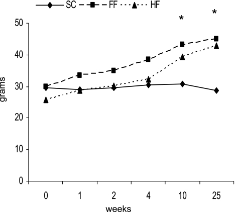
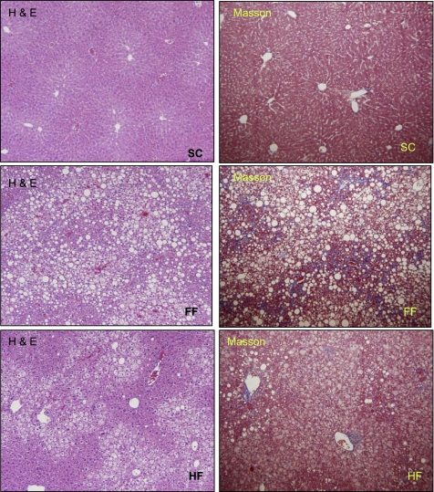
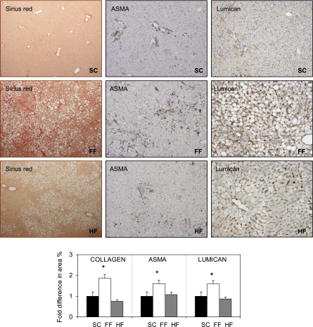
Although there are small animal platforms that recapitulate some of the histological features of nonalcoholic fatty liver disease, there are no small animal models of nonalcoholic steatohepatitis (NASH) with consistent hepatocellular ballooning and progressive fibrosis that also exhibit fidelity to the human condition physiologically. We examined the metabolic and histological effects of a diet on the basis of the composition of "fast food" (high saturated fats, cholesterol, and fructose). Mice (n = 8 in each group) were assigned to diets as follows: 1) standard chow (SC), i.e., 13% energy as fat [1% saturated fatty acids (SFA)], 2) high fat (HF), i.e., 60% energy as fat (1% SFA), and 3) fast food (FF), i.e., 40% energy as fat (12% SFA, 2% cholesterol). All three diets were supplemented with high fructose. All diets produced obesity. The HF and FF diets produced insulin resistance. Liver histology was normal in animals fed the SC diet. Steatohepatitis with pronounced ballooning and progressive fibrosis (stage 2) was observed in mice fed the FF diet. Although the HF diet produced obesity, insulin resistance, and some steatosis; inflammation was minimal, and there was no increase in fibrosis. The FF diet produced a gene expression signature of increased fibrosis, inflammation, and endoplasmic reticulum stress and lipoapoptosis. A diet based on high cholesterol, high saturated fat, and high fructose recapitulates features of the metabolic syndrome and NASH with progressive fibrosis. This represents a novel small animal model of fibrosing NASH with high fidelity to the human condition. These results highlight the contribution of dietary composition to the development of nonalcoholic fatty liver disease and NASH.
Figures

References
-
- Adams LA, Lymp JF, St Sauver, Sanderson SO, Lindor KD, Feldstein A, Angulo P. The natural history of nonalcoholic fatty liver disease: a population-based cohort study. Gastroenterology 129: 113–121, 2005 - PubMed
-
- Angulo P. Nonalcoholic fatty liver disease and liver transplantation. Liver Transpl 12: 523–534, 2006 - PubMed
-
- Angulo P. Nonalcoholic fatty liver disease. J Gastroenterol Hepatol 346: 1221–1231, 2002 - PubMed
-
- Angulo P, Alba LM, Petrovic LM, Adams LA, Lindor KD, Jensen MD. Leptin, insulin resistance, and liver fibrosis in human nonalcoholic fatty liver disease. J Hepatol 41: 943–949, 2004 - PubMed
Publication types
MeSH terms
Grants and funding
LinkOut - more resources
Full Text Sources
Other Literature Sources
Medical
Research Materials
Miscellaneous