Comparative Study
. 2011 Nov;31(11):2749-59.
doi: 10.1161/ATVBAHA.111.235176.

Activation of the annexin A1 pathway underlies the protective effects exerted by estrogen in polymorphonuclear leukocytes

Affiliations
Comparative Study

Activation of the annexin A1 pathway underlies the protective effects exerted by estrogen in polymorphonuclear leukocytes

Suchita Nadkarni et al. Arterioscler Thromb Vasc Biol. 2011 Nov.

Abstract

Objective: The anti-inflammatory properties of the female sex hormone estrogen have been linked to a reduced incidence of cardiovascular disease. In the present study, we addressed whether estrogen could activate vasculoprotective mechanisms via annexin A1 (AnxA1) mobilization in human polymorphonuclear cells (PMNs).

Methods and results: Using whole-blood flow cytometry, we demonstrated that premenopausal women expressed higher levels of surface AnxA1 on circulating PMNs compared with males. This correlated with high plasma estrogen during the menstrual cycle. The addition of estrogen in vitro to male PMNs induced rapid mobilization of AnxA1, optimal at 5 ng/mL and a 30-minute incubation period; this effect was abolished in the presence of the estrogen receptor antagonist ICI182780. Estrogen addition to human PMNs induced a distinct AnxA1(hi) CD62L(lo) CD11b(lo) phenotype, and this was associated with lower cell activation as measured by microparticle formation. Treatment of human PMNs with E(2) inhibited cell adhesion to an endothelial cell monolayer under shear, which was absent when endogenous AnxA1 was neutralized. Of interest, addition of estrogen to PMNs flowed over the endothelial monolayer amplified its upregulation of AnxA1 localization on the cell surface. Finally, in a model of intravital microscopy, estrogen inhibition of white blood cell adhesion to the postcapillary venule was absent in mice nullified for AnxA1.

Conclusion: We unveil a novel AnxA1-dependent mechanism behind the inhibitory properties of estrogen on PMN activation, describing a novel phenotype with a conceivable impact on the vasculoprotective effects of this hormone.


Figures

Figure 1
Figure 1. Gender differences in AnxA1 mobilization on PMN surface
A) Different degree of expression of AnxA1 on PMN cell surface between males and females. Freshly prepared cells were stained and gated on CD16+ve PMN, then double stained for AnxA1 and FPR2/ALX. Data report the % of positive cells, and are mean ± SEM of 6 subjects per group. * P<0.05 (Student’s t Test). B) Dose-response and time-course of E2-mediated mobilization of AnxA1 on the PMN cell surface. Healthy male blood was incubated at 37°C with the indicated concentrations of E2 for 30 min (left graph), or at 5 ng/ml E2 for different times (right graph). Cells were gated as described in panel A. Data are mean ± SEM of 5-6 analyses with distinct cell preparations. **P<0.001 (One-way ANOVA with Dunnett’s post-correction [left graph] or two-way ANOVA with Bonferroni post correction [right graph]). C) Western blotting analysis for AnxA1 expression in PMN cytosolic and membrane fractions in the absence or presence of E2 (5ng/ml; 30 min). Blots are representative of three experiments. D) Involvement of ER in E2-induced AnxA1 mobilization. Whole blood aliquots were pre-incubated for 10 min at 37°C with the non-specific ER antagonist ICI182780, followed by a 30-min incubation with E2 (5 ng/ml). AnxA1 mobilization was analysed by flow cytometry as in panel A. Data are mean ± SEM of 5 experiments. *P<0.001 compared to vehicle without E2 (veh); # P< 0.001 compared to vehicle + E2 (two-way ANOVA with Bonferroni post correction).
Figure 2
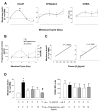
Figure 2. AnxA1 mobilization on PMN in females varies during the menstrual cycle
A) Fluctuations in AnxA1, FPR2/ALX and CD62L cell surface expression of circulating PMN from healthy females on day 2, 12, 19, and 26 of their menstrual cycle. Data are mean ± SEM of 6 subjects; **P<0.001 vs. day 2 (two-way ANOVA with Bonferroni post correction). Dotted line indicates values obtained in cells from male volunteers. B) Plasma levels of estrogen, progesterone and cortisol during the menstrual cycle. Time points as in Panel A. C) Correlation between circulating estrogen levels and AnxA1 expression on the PMN cell surface. Estrogen levels were correlated with AnxA1+ve PMN (r2=0.5628; left graph). Exclusion of Day 26 values, denoted by arrows, yields the graph on the right with a calculated r2 = 0.7655. D) Progesterone counteracts the effect of estrogen on AnxA1. Progesterone (5 and 10 ng/ml) was added to whole blood in the absence or presence of E2 (5 ng/ml) for a 30 min incubation period. The degree of AnxA1 and CD62L expression on the PMN was quantified by flow cytometry. Data are mean ± SEM of three distinct experiments. *P<0.05 vs. control (dose 0 group); #P<0.05 vs. E2 alone (one-way ANOVA with Dunnett’s post correction).
Figure 3
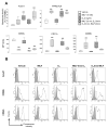
Figure 3. Estrogen confers an anti-inflammatory phenotype to human PMN
A) Male whole blood was treated with either fMLF (10 nM) for 20 min, E2 (5 ng/ml) for 30 min, or a combination of treatments, that is fMLF, followed by E2 or pre-treatment with E2 followed by fMLF. All blood aliquots were kept at 37°C for a total 50 min incubation time. Flow cytometry afforded quantification of the degree of expression of AnxA1, FPR2/ALX, CD62L, CD11b, and CD66b on PMN cell surface. Data are mean ± SEM of 6 experiments conducted with distinct blood preparations. *P<0.001 compared to vehicle control (one-way ANOVA with Dunnett’s post correction). B) Representative histograms depicting the mean fluorescence units (MFI) for AnxA1, CD66b and CD62L out of the experiments presented in Panel A. Grey histograms indicate isotype controls (IgG1κ for AnxA1 and CD62L, and IgMκ for CD66b).
Figure 4
Figure 4. Release of PMN-derived microparticles in plasma is regulated by estrogen
Plasma was collected from blood treated as described in Figure 4. A) PMN microparticles in the plasma were identified with CD66b staining and calculated as percentage of the total microparticles. Data are mean ± SEM of 6 experiments conducted with distinct blood preparations. *P<0.001compared to vehicle control (one-way ANOVA with Dunnett’s post correction). B) Representative histogram plots for CD66b in relation to the different treatments. Grey histograms indicate isotype control (mouse IgMκ). C) Further characterization of CD66b+ve microparticles with double staining for AnxA1, FPR2/ALX and CD62L, reporting variation in mean fluorescence intensity (MFI) units for each antigen. Data are mean ± SEM of 6 experiments conducted with distinct blood preparations. *P<0.001 compared to vehicle control (one-way ANOVA with Dunnett’s post correction).
Figure 5
Figure 5. E2 inhibits PMN adhesion to inflamed endothelium in an AnxA1-dependent fashion
Human umbilical vein endothelial cells (HUVEC) were stimulated with TNF-α (10ng/ml) for 4 hours. Human PMN were treated with either vehice, E2 (5ng/ml) or with E2 plus AnxA1 neutralizing Ab (20μg/ml) for 30 min. PMN were then flowed over the stimulated endothelial monolayer for 8 min, after which 6 random fields were recorded. A) PMN capture, adhesion and rolling were measured. *P< 0.001 E2 compared to vehicle. # P< 0.001 E2 compared to E2 plus AnxA1 neutralizing Ab. B) Representative images of PMN interactions with endothelium under flow. Following analysis using Image-Pro, neutrophils that have firmly adhered to the endothelium are situated within the dotted circles. Empty dotted circles indicate non-adhered neutrophils.
Figure 6
Figure 6. E2- treated PMN post flow have a marked increase in AnxA1 surface expression, but low CD11b
PMN were treated as described in Figure 5. Pre-flow (A), post-flow (B) and post-adherent PMN were stained for AnxA1, CD62L and CD11b expression on the plasma membrane. P< 0.001 compared to vehicle (Student’s T test). Representative histograms are also shown.

Similar articles

Cited by

References

    1. Kannel WB, Wilson P. Risk factors that attenuate the female coronary disease advantage. Arch Intern Med. 1995;155:57–61. - PubMed
    1. Bakir S, Mori T, Durand J, Chen YF, Oparil S. Estrogen-induced vasoprotection is estrogen receptor dependent: evidence from the balloon-injured rat carotid artery mode. Circulation. 2000;101:2342–2344. - PubMed
    1. Chen SJ, Li H, Durand J, Oparil S, Chen Y. Estrogen reduces myointimal proliferation after balloon injury of rat carotid artery. Circulation. 1996;93:577–584. - PubMed
    1. Sherwood A, Bower JK, McFetridge-Durdle J, Blumenthal JA, Newby LK, Hinderliter A. Age moderates the short-term effects of transdermal 17beta-estradiol on endothelium-dependent vascular function in postmenopausal women. Arterioscler Thromb Vasc. 2007;27:1782–1787. - PubMed
    1. Hayashi T, Yamada K, Esaki T, Kuzuya M, Satake S, Ishikawa T, Hidaka H, Iguchi A. Estrogen increases endothelial nitric oxide by a receptor-mediated system. Biochem Biophys. 1995;214:847–855. - PubMed

Publication types

MeSH terms