

Late occurrence of granular dystrophy in bilateral keratoconus: penetrating keratoplasty and long-term follow-up
- PMID: 21836353
- PMCID: PMC3159329
- DOI: 10.4103/0301-4738.83624
Late occurrence of granular dystrophy in bilateral keratoconus: penetrating keratoplasty and long-term follow-up
Abstract
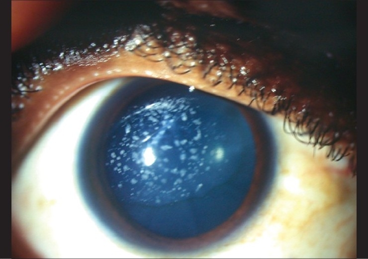
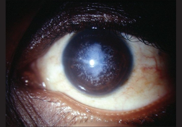
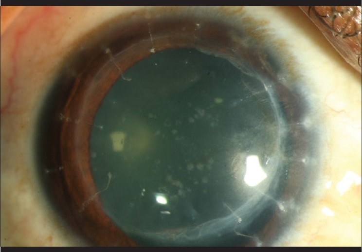
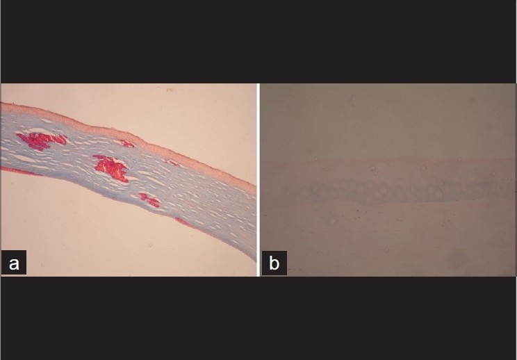
We report a rare case of keratoconus with granular dystrophy with a follow-up of two decades, documenting the sequential presentation of two diseases confirmed by histology and genetic studies. A 13-year-old boy was diagnosed in 1988 with keratoconus in both eyes (BE) based on slit-lamp biomicroscopy findings of corneal ectasia in BE accompanied by Fleischer's ring, Vogt's striae, a small, old, healed hydrops. The left eye (LE) had central corneal thinning and scar in the central area involving the mid and posterior stroma secondary to healed hydrops. Penetrating keratoplasty (PKP) was advised. The boy was lost to follow-up till 1991 and presented with white, dot-like opacities in the central cornea in the RE only, suggestive of granular corneal dystrophy. Similar findings of white, dot-like opacities were noted in the LE in 1995 and the patient subsequently underwent PKP in BE. Histopathology of corneal buttons confirmed the presence of patchy, crystal-like orange deposits, which stained bright red with Masson's trichrome. Mutational analysis of the TGFBI gene in patient's DNA revealed a heterozygous mutation corresponding to a change in Arg555Trp in the keratoepithelin protein. Granular dystrophy recurred after 8 years in the RE.
Figures
Similar articles
-
Bilateral Atypical Granular Corneal Dystrophy Associated with Unilateral Keratoconus in a Male Child.Middle East Afr J Ophthalmol. 2016 Jul-Sep;23(3):262-4. doi: 10.4103/0974-9233.186151. Middle East Afr J Ophthalmol. 2016. PMID: 27555713 Free PMC article.
-
Concurrent macular corneal dystrophy and keratoconus.Middle East Afr J Ophthalmol. 2012 Apr-Jun;19(2):251-3. doi: 10.4103/0974-9233.95266. Middle East Afr J Ophthalmol. 2012. PMID: 22623870 Free PMC article.
-
A case of concomitant keratoconus and granular corneal dystrophy type II.Cont Lens Anterior Eye. 2014 Aug;37(4):314-6. doi: 10.1016/j.clae.2014.02.001. Epub 2014 Feb 28. Cont Lens Anterior Eye. 2014. PMID: 24582869
-
Stage-related therapy of corneal dystrophies.Dev Ophthalmol. 2011;48:116-153. doi: 10.1159/000324081. Epub 2011 Apr 26. Dev Ophthalmol. 2011. PMID: 21540634 Review.
-
Keratoconus.Optom Clin. 1995;4(3):65-73. Optom Clin. 1995. PMID: 7767020 Review.
Cited by
-
Familial association of keratoconus and granular corneal dystrophy: The familial case series.North Clin Istanb. 2018 Sep 4;6(2):176-183. doi: 10.14744/nci.2018.08860. eCollection 2019. North Clin Istanb. 2018. PMID: 31297486 Free PMC article.
-
Bilateral Atypical Granular Corneal Dystrophy Associated with Unilateral Keratoconus in a Male Child.Middle East Afr J Ophthalmol. 2016 Jul-Sep;23(3):262-4. doi: 10.4103/0974-9233.186151. Middle East Afr J Ophthalmol. 2016. PMID: 27555713 Free PMC article.
-
Systematically Displaying the Pathogenesis of Keratoconus via Multi-Level Related Gene Enrichment-Based Review.Front Med (Lausanne). 2022 Jan 24;8:770138. doi: 10.3389/fmed.2021.770138. eCollection 2021. Front Med (Lausanne). 2022. PMID: 35141241 Free PMC article.
References
-
- Rabinowitz YS. Keratoconus. Surv Ophthalmol. 1998;42:297–319. - PubMed
-
- Sassani JW, Smith SG, Rabinowitz YS. Keratoconus and bilateral lattice-granular corneal dystrophies. Cornea. 1992;11:343–50. - PubMed
-
- Vajpayee RB, Snibson GR, Taylor HR. Association of keratoconus with granular corneal dystrophy. Aust N Z J Ophthalmol. 1996;24:369–71. - PubMed
-
- Wollensak G, Green WR, Temprano J. Keratoconus associated with corneal granular dystrophy in a patient of Italian origin. Cornea. 2002;21:121–2. - PubMed
-
- Cremona FA, Ghoshesh FR, Rapuano CJ, Eagle RC, Jr, Hammersmith KM, Laibson PR, et al. Keratoconus associated with other corneal dystrophies. Cornea. 2009;28:127–35. - PubMed
Publication types
MeSH terms
LinkOut - more resources
Full Text Sources
Miscellaneous
