. 2011 Sep;35(9):1311-26.
doi: 10.1097/PAS.0b013e31821ec4ce.

Primary bimorphic adrenocortical disease: cause of hypercortisolism in McCune-Albright syndrome

Affiliations

Primary bimorphic adrenocortical disease: cause of hypercortisolism in McCune-Albright syndrome

J Aidan Carney et al. Am J Surg Pathol. 2011 Sep.

Abstract

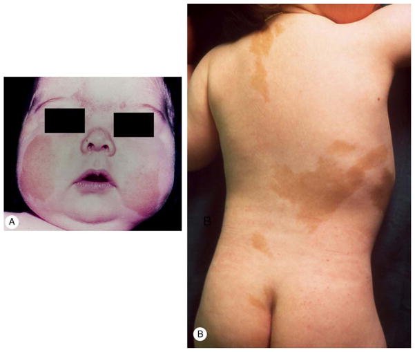
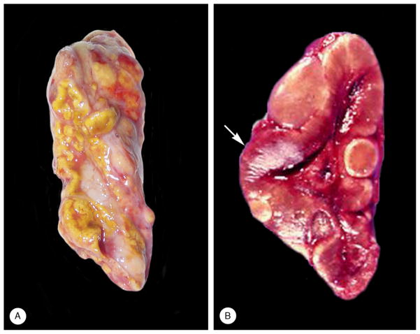
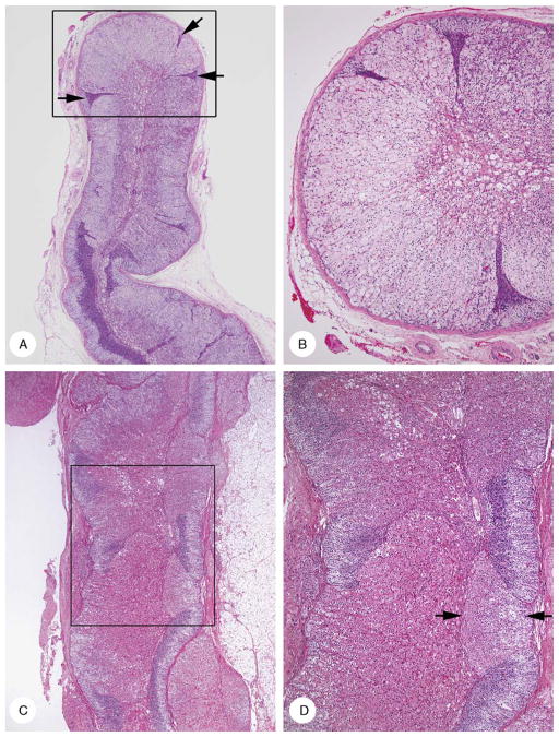
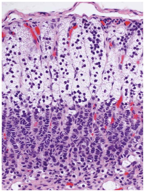
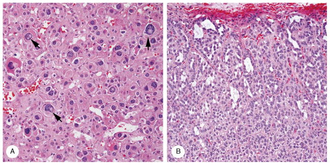
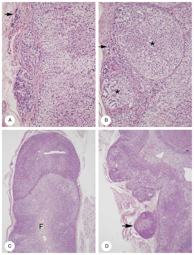
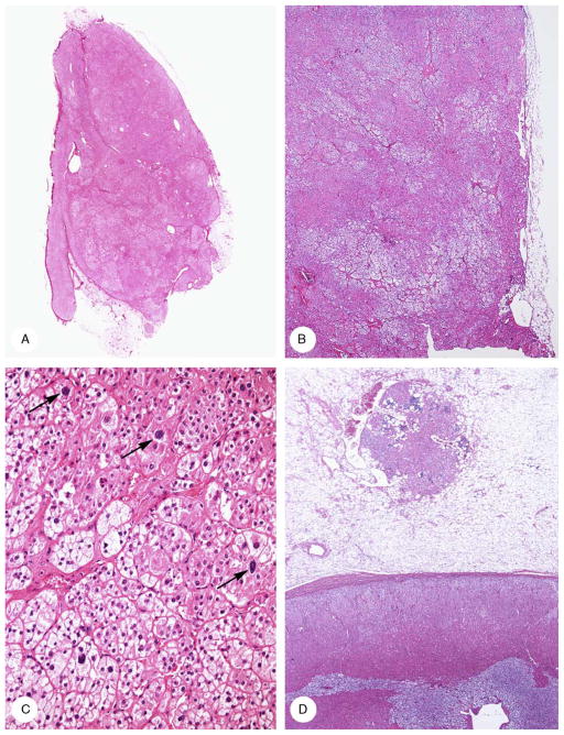
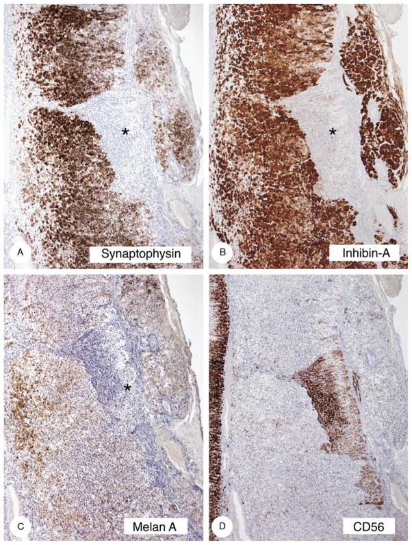
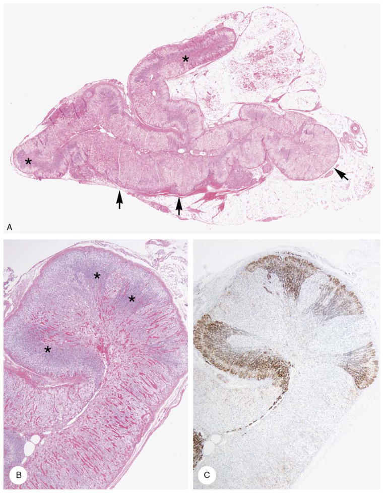
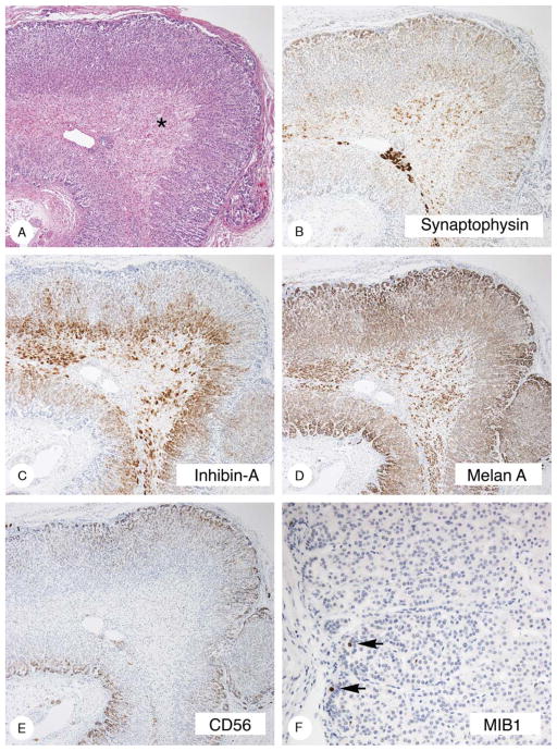
McCune-Albright syndrome (polyostotic fibrous dysplasia, café-au-lait skin spots, and precocious puberty) is a genetically mosaic disorder with populations of mutant and normal cells in affected organs. Cushing syndrome, a rare feature of the condition, usually affects infants and is the result of corticotropin-independent primary bilateral adrenal disease, usually interpreted as nodular adrenocortical hyperplasia. In this study of 9 patients with Cushing syndrome and McCune-Albright syndrome, light microscopy revealed a characteristic bimorphic pattern of diffuse and nodular hyperplasia and a distinctive form of cortical atrophy with apparent zona glomerulosa hyperplasia in 8 patients, all very young. The pattern could be explained by the presence of a mosaic distribution of mutant and normal cells in the adrenal glands. The findings are different from those in inherited or other forms of genetically caused Cushing syndrome. The ninth patient, aged 17 years, had an adrenal adenoma and diffuse cortical hyperplasia in each adrenal gland.


Conflict of interest statement

Conflicts of Interest and Sources of Funding: J.A.C. received funding from the Mayo Foundation and the Intramural Program, NIH project Z01HD00064204.

Figures

FIGURE 1
FIGURE 1
A, Case 7. Moon-face and facial plethora in a 4-month-old male infant. B, Case 9. Multiple café-au-lait patches in a 2-month-old female infant.
FIGURE 2
FIGURE 2
Gross appearance of adrenal gland [case 3 (A) and case 5 (B)]. A, The external surface was yellow and partially covered with fat. Several nodules were visible. B, The reddish-brown thickened cortex (arrow) featured several “tan-to-green nodules.”
FIGURE 3
FIGURE 3
Cortical hyperplasia and cortical atrophy [A and B (case 7); C and D (case 8)]. A, Low-power micrograph showed diffuse clear cell hyperplasia with variable thickening of the cortex. Hyperplasia was punctuated by compressed and stretched zones of atrophy (arrows). B, Intermediate-power magnification area in box in panel A. The cortex lacked zonation and was composed of hyperplastic clear cells punctuated by 3 narrow tongues of atrophic cortex. Fetal cortex with vacuolar degeneration was present centrally. C, Cortex had a variegated appearance due to alternating areas of acidophilic cell hyperplasia interspersed in areas of cortical atrophy (outer light and inner dark layers). D, Intermediate-power magnification of area in box in panel C. Compact cell hyperplasia had 2 tinctorial appearances; moderately strong (arrows) and more intense (lower center).
FIGURE 4
FIGURE 4
Cortical atrophy (case 7). Cortex was 0.3-mm thick. There was a subcapsular layer of vacuolated cells with round dense nuclei and inner layer of rows of cells with molded nuclei and scarcely visible cytoplasm.
FIGURE 5
FIGURE 5
Cortical cytomegaly and microcysts (case 1). A, Cytomegaly featured large cells with eosinophilic cytoplasm and large to huge nuclei, several with intranuclear vacuoles (arrows). B, A series of polymorphic spaces (microcysts) separate the 2-cell columns of the subcapsular cortex.
FIGURE 6
FIGURE 6
Patterns of adrenal capsular involvement (case 6). A, Three intracapsular aggregates, 1 very small (arrow). The cytology and pattern of the cells are different in each of the aggregates. B, The capsule is distended by 2 nodules (asterisks), each having a thin fibrous capsule derived from the original capsule. In addition, rows of cortical cells are present in the capsule (arrow). The cells in the larger nodule (upper right) resemble those in the cortex proper (bottom right). Some of the cells in the smaller nodule (lower left) resemble those in the linear rows in the capsule. C, A roughly hemispherical mass of cortical cells with a very thin capsule is applied to the tip of a cortical ala in which the cortex is hyperplastic and lacks zonation. Fetal cortex is present at the center of the gland (F). D, A series of partially encapsulated nodules caused thickening of the cortex. The latter showed vague zonation, an outer clear zone with clear cytoplasm, and an inner acidophilic one with acidophilic cytoplasm (bottom half). The thickening of the cortex is increased by a series of partially encapsulated nodules (upper half). One nodule (arrow) is close to but separate from the adrenal gland. There is no infiltration of periadrenal fat.
FIGURE 7
FIGURE 7
Bilateral adrenocortical adenoma (case 4). A, A circumscribed, lobulated tumor (autopsy specimen) protruded from the hyperplastic right adrenal cortex that lacked normal zonation. B, The left adrenal tumor (surgical specimen) was composed of acidophilic and clear cells and lacked a capsule. C, Tumor was composed of a sheet of cells with eosinophilic cells (upper right) and clustered clear cells (lower left). Scattered nuclei were enlarged (arrows). D, An unencapsulated 1.5-mm nodule of adrenocortical cells with eosinophilic cytoplasm and lymphocytic infiltrates in the periadrenal fat. The region of the zona fasciculata in the cortex proper lacked the normal clear appearance.
FIGURE 8
FIGURE 8
Immunostaining of hyperplastic and atrophic cortex (case 8). The hyperplastic cortex stained strongly with synaptophysin (A) and inhibin-A (B), less strongly with melan A (C), and did not stain or stained minimally with CD56 (D). The atrophic cortex (asterisks) stained strongly with CD56 and minimally with melan A.
FIGURE 9
FIGURE 9
Cortical hyperplasia and cortical atrophy (case 10). A, Areas of hyperplasia with mixed clear and acidophilic cells (arrows) are juxtaposed with zones of atrophic cortex with outer clear and inner dark cells (asterisks). B, Mixed clear and acidophilic cell hyperplasia with cortical atrophy (arrows). C, CD56-stained zones of cortical atrophy. Zones of mixed clear and acidophilic cell hyperplasia were unstained.
FIGURE 10
FIGURE 10
Definitive and residual fetal adrenal cortex (3-month-old male infant). A, Definitive cortex lacked distinct zonation, was 0.5-mm thick, and surrounded the fetal cortex (asterisk). B, Synaptophysin stained superficial and deep bands of the definitive cortex leaving a central zone unstained. Medullary cells were heavily stained. Fetal cortical cells were less heavily stained. C, Inhibin-A stained a band of deep definitive cortical and fetal cells. D, Melan A stained the entire cortex and also fetal cortical cells. E, CD56 stained a continuous subcapsular band of cortical cells. F, MIB1 stained scattered nuclei in the atrophic cortex (arrows).

References

    1. Aarskog D, Tveteraas E. McCune-Albright’s syndrome following adrenalectomy for Cushing syndrome in infancy. J Pediatr. 1968;73:89–96. - PubMed
    1. Albright F, Butler AM, Bloomberg E. Rickets resistant to vitamin D therapy. Am J Dis Child. 1937;54:529–547.
    1. Albright F, Butler AM, Hampton AO, et al. Syndrome characterized by osteitis fibrosa disseminata, areas of pigmentation and endocrine dysfunction, with precocious puberty in females: report of five cases. N Engl J Med. 1937;216:727–746.
    1. Beckwith JB. Macroglossia, omphalocele, adrenal cytomegaly, gigantism, and hyperplastic visceromegaly. Birth Defects. 1969;5:188–196.
    1. Benjamin DR, McRoberts JW. Polyostotic fibrous dysplasia associated with Cushing syndrome. Arch Pathol. 1973;96:175–178. - PubMed

MeSH terms