

Optic disc classification by the Heidelberg Retina Tomograph and by physicians with varying experience of glaucoma
- PMID: 21836629
- PMCID: PMC3213643
- DOI: 10.1038/eye.2011.172
Optic disc classification by the Heidelberg Retina Tomograph and by physicians with varying experience of glaucoma
Abstract
Purpose: To compare the diagnostic accuracy of the Heidelberg Retina Tomograph's (HRT) Moorfields regression analysis (MRA) and glaucoma probability score (GPS) with that of subjective grading of optic disc photographs performed by ophthalmologists with varying experience of glaucoma and by ophthalmology residents.
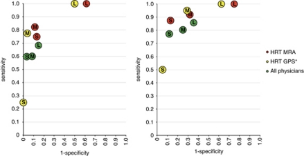
Methods: Digitized disc photographs and HRT images from 97 glaucoma patients with visual field defects and 138 healthy individuals were classified as either within normal limits (WNL), borderline (BL), or outside normal limits (ONL). Sensitivity and specificity were compared for MRA, GPS, and the physicians. Analyses were also made according to disc size and for advanced visual field loss.
Results: Forty-five physicians participated. When BL results were regarded as normal, sensitivity was significantly higher (P<5%) for both MRA and GPS compared with the average physician, 87%, 79%, and 62%, respectively. Specificity ranged from 86% for MRA to 97% for general ophthalmologists, but the differences were not significant. In eyes with small discs, sensitivity was 75% for MRA, 60% for the average doctor, and 25% for GPS; in eyes with large discs, sensitivity was 100% for both GPS and MRA, but only 68% for physicians.
Conclusion: Our results suggest that sensitivity of MRA is superior to that of the average physician, but not that of glaucoma experts. MRA correctly classified all eyes with advanced glaucoma and showed the best sensitivity in eyes with small optic discs.
Figures
References
-
- Morgan JE, Sheen NJ, North RV, Goyal R, Morgan S, Ansari E, et al. Discrimination of glaucomatous optic neuropathy by digital stereoscopic analysis. Ophthalmology. 2005;112:855–862. - PubMed
-
- Wollstein G, Garway-Heath DF, Fontana L, Hitchings RA. Identifying early glaucomatous changes. Comparison between expert clinical assessment of optic disc photographs and confocal scanning ophthalmoscopy. Ophthalmology. 2000;107:2272–2277. - PubMed
Publication types
MeSH terms
LinkOut - more resources
Full Text Sources
