. 2011 Sep 30;12(10):1069-76.
doi: 10.1038/embor.2011.151.

CREB and ChREBP oppositely regulate SIRT1 expression in response to energy availability

Affiliations

CREB and ChREBP oppositely regulate SIRT1 expression in response to energy availability

Lilia G Noriega et al. EMBO Rep. .

Abstract

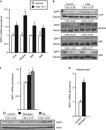
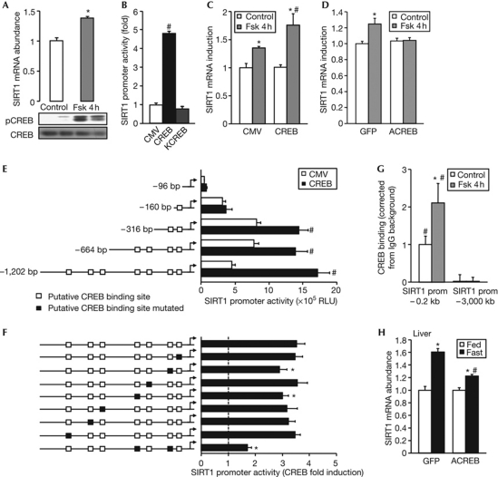
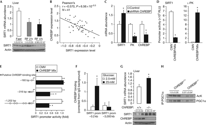
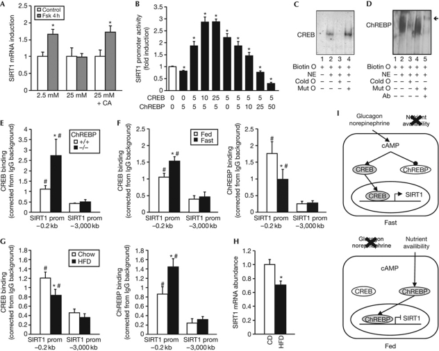
The nicotinamide adenine dinucleotide (NAD(+))-dependent deacetylase SIRT1 is a major metabolic regulator activated by energy stresses such as fasting or calorie restriction. SIRT1 activation during fasting not only relies on the increase in the NAD(+)/NADH ratio caused by energy deprivation but also involves an upregulation of SIRT1 mRNA and protein levels in various metabolic tissues. We demonstrate that SIRT1 expression is controlled systemically by the activation of the cyclic AMP response-element-binding protein upon low nutrient availability. Conversely, in the absence of energetic stress, the carbohydrate response-element-binding protein represses the expression of SIRT1. Altogether, these results demonstrate that SIRT1 expression is tightly controlled at the transcriptional level by nutrient availability and further underscore that SIRT1 is a crucial metabolic checkpoint connecting the energetic status with transcriptional programmes.


Conflict of interest statement

The authors declare that they have no conflict of interest.

Figures

Figure 1
Figure 1
SIRT1 expression is induced by low nutrient availability and in response to humoral factors released during fasting. (A) SIRT1 mRNA levels in the liver, gastrocnemius muscle, BAT and epididymal WAT of mice fasted for 12 h. The control group had free access to food (n=4). (B) SIRT1 protein levels in the liver, gastrocnemius, BAT and epididymal WAT of control or 24 h-fasted mice. (C) SIRT1 mRNA and protein levels in the liver of mice 1 h after the intraperitoneal administration of PBS (control), glucagon (50 μg/kg) or NE (1 mg/kg; n=5). (D) Quantitative PCR analysis of SIRT1 mRNA levels in primary mouse hepatocytes stimulated for 2 h with 100 nM glucagon (n=4). Protein levels were measured by western blot using tubulin or actin as loading control. Values are presented as the average±s.e.m. and asterisk indicates a statistical difference compared with control (P-value <0.05). BAT, brown adipose tissue; NE, norepinephrine; WAT, white adipose tissue.
Figure 2
Figure 2
SIRT1 expression is transcriptionally activated by CREB. (A) SIRT1 mRNA in HepG2 cells stimulated for 4 h with 10 μM Fsk (n=4). pCREB and CREB protein levels were analysed by western blot analysis. (B) HepG2 cells were transfected with a SIRT1 promoter luciferase reporter, a β-galactosidase normalization plasmid and 10 ng of pCMV–CREB or pCMV–KCREB. Luciferase activity was measured 48 h after transfection and values were normalized to β-galactosidase levels (n=4). Values are presented as fold increase over the empty vector (CMV; n=4). SIRT1 mRNA levels in (C) HepG2 cells transfected with pCMV–CREB or an empty vector (CMV; n=4) and (D) HepG2 infected with adenoviruses encoding GFP or the dominant-negative ACREB (n=6), after 4 h stimulation with 10 μM Fsk. Data are relative to control group. HepG2 cells were transfected with either (E) 10 ng of pCMV–CREB and serial deletions of the SIRT1 promoter luciferase reporter or (F) the SIRT1 promoter luciferase reporter containing site-directed mutagenesis of the indicated putative CREB-binding site. Luciferase activity was measured 48 h after transfection. (G) HepG2 cells were treated for 4 h with Fsk, and ChIP was performed using an IgG control or a CREB-specific antibody. Recruitment of CREB to the SIRT1 promoter was measured by qPCR using primers against the proximal −0.2 kb and the distal −3 kb promoter, and corrected for background measured in IgG immunoprecipitates (n=4). (H) SIRT1 mRNA abundance in liver from fed or 18 h-fasted mice infected with adenovirus expressing GFP or ACREB. Values are presented as the average±s.e.m., asterisk indicates statistical difference compared with control (A,C,D), WT SIRT1 promoter luciferase reporter (F) or fed group (G) and hash symbol indicates statistical difference compared with empty vector (B,E), distal promoter (G) or GFP (H) at P<0.05. ChIP, chromatin immunoprecipitation; CMV, cytomegalovirus; CREB, cyclic AMP response-element-binding protein; Fsk, forskolin; GFP, green fluorescent protein; IgG, immunoglobulin G; pCMV, CMV promoter; pCREB, CREB phosphorylation; prom, promoter; qPCR, quantitative PCR; RLU, relative luciferase units; WT, wild type.
Figure 3
Figure 3
ChREBP transcriptionally represses SIRT1 expression. (A) SIRT1 mRNA and protein levels in the liver of mice fasted for 24 h or refed for 2 or 4 h. (B) Correlation plots for the liver mRNA expression of SIRT1 and ChREBP in the mouse BxD genetic reference population. (C) SIRT1, L-PK and ChREBP mRNA levels in HepG2 cells transfected with a ChREBP shRNA (n=4). (D) HepG2 cells were transfected with a SIRT1 or L-PK promoter luciferase reporter, β-galactosidase, and 100 ng of an empty vector (CMV) or 50 ng of ChREBP and 50 ng of Mlx expression vectors. Luciferase activity was measured as described (n=4). (E) HepG2 cells were transfected with −1,202, −316 or −160 bp fragment of the SIRT1 promoter luciferase reporter and ChREBP and Mlx plasmid. Luciferase activity was measured 48 h after transfection and values were normalized to total protein (n=6). Values are presented as fold increase over the empty vector (CMV). (F) HepG2 cells were incubated 20 h with 2.5 or 25 mM glucose; further, ChIP was performed using an IgG control or a ChREBP-specific antibody. Recruitment of ChREBP to the SIRT1 promoter was measured as described (n=4). (G) SIRT1 mRNA and protein levels in liver lysates of fed ChREBP+/+ or ChREBP−/− mice. Actin was used as loading control. (H) PGC1α acetylation levels were examined in liver from fed ChREBP+/+ or ChREBP−/− mice after immunoprecipitation. The quantification represents the ratio of acetylated to total PGC1α. Values are the fold induction over control/empty vector and are presented as the average±s.e.m., and asterisk indicates a statistical difference compared with fast/control/empty vector/ChREBP+/+ or 2.5 mM glucose at P<0.05. Ack, acetylated lysine; ChIP, chromatin immunoprecipitation; ChREBP, carbohydrate response-element-binding protein; CMV, cytomegalovirus; IP-PGC1α, immunoprecipitated PGC1α; IgG, immunoglobulin G; PGC1α, peroxisome proliferator-activated receptor gamma coactivator 1α; PK, pyruvate kinase; prom, promoter; RF, refed; RLU, relative luciferase units; shRNA, short hairpin RNA.
Figure 4
Figure 4
The regulation of SIRT1 by CREB and ChREBP is interdependent and coordinated by energy availability. (A) HepG2 cells were stimulated for 4 h with 10 μM forskolin in 2.5 mM or 25 mM glucose. A third group was treated with CA 1 h before treatment of forskolin in 25 mM glucose. SIRT1 mRNA levels were then measured by qPCR. Data are relative to the control group. (B) HepG2 cells were transfected with the SIRT1 promoter luciferase reporter and with the indicated amount (ng) of pCMV–CREB and ChREBP/Mlx. Luciferase activity was measured as described (n=6). (C) EMSA was performed using nuclear extracts from CHO cells transfected with CREB and a 25-bp biotinylated oligonucleotide with the overlapping core sequence for CREB and ChREBP. (D) EMSA was performed as in C, using nuclear extracts from CHO cells transfected with Flag-tagged ChREBP and its partner Mlx. Arrow indicates the supershifted band. (E) ChIP was performed in the liver from fed ChREBP+/+ or ChREBP−/− mice using an IgG control or CREB-specific antibody. (F) ChIP was performed in the liver from fed or 18 h-fasted mice using an IgG control, CREB- or ChREBP-specific antibody. (G) ChIP was performed in the liver from mice fed chow or HFD using an IgG control or CREB- or ChREBP-specific antibody as described previously. (H) SIRT1 mRNA abundance in the liver of mice fed chow or HFD. Values are presented as the average±s.e.m., asterisk indicates a statistical difference compared with control/empty vector/ChREBP+/+/FED or chow, and hash symbol indicates statistical difference compared with distal promoter at P<0.05. (I) Scheme illustrating how nutrient availability is integrated by CREB and ChREBP to coordinately regulate SIRT1 expression. During fasting, glucagon and norepinephrine lead to an increase in cAMP and PKA activity, in turn leading to CREB activation and ChREBP inactivation. CREB then induces SIRT1 expression. Conversely, during the fed state, ChREBP binds to the SIRT1 promoter to downregulate its expression, potentially through competition with CREB binding (bottom panel). Ab, flag-specific antibody; biotin O, biotinylated oligonucleotide; CA, cantharidic acid; cAMP, cyclic AMP; ChIP, chromatin immunoprecipitation; ChREBP, carbohydrate response-element-binding protein; cold O, cold oligonucleotide excess; CREB, cAMP response-element-binding protein; EMSA, electrophoretic mobility shift assay; Fsk, forskolin; HFD, high-fat diet; IgG, immunoglobulin G; mut O, cold mutant oligonucleotide excess; NE, nuclear extract; pCMV, CMV promoter; PKA, protein kinase A; prom, promoter; qPCR, quantitative PCR.

Comment in

  • Metabolic signals regulate SIRT1 expression.
    Chalkiadaki A, Guarente L. Chalkiadaki A, et al. EMBO Rep. 2011 Sep 30;12(10):985-6. doi: 10.1038/embor.2011.179. EMBO Rep. 2011. PMID: 21941300 Free PMC article. No abstract available.

References

    1. Berry MN, Friend DS (1969) High-yield preparation of isolated rat liver parenchymal cells: a biochemical and fine structural study. J Cell Biol 43: 506–520 - PMC - PubMed
    1. Brunet A et al. (2004) Stress-dependent regulation of FOXO transcription factors by the SIRT1 deacetylase. Science 303: 2011–2015 - PubMed
    1. Burgess SC, Iizuka K, Jeoung NH, Harris RA, Kashiwaya Y, Veech RL, Kitazume T, Uyeda K (2008) Carbohydrate-response element-binding protein deletion alters substrate utilization producing an energy-deficient liver. J Biol Chem 283: 1670–1678 - PubMed
    1. Canto C, Jiang LQ, Deshmukh AS, Mataki C, Coste A, Lagouge M, Zierath JR, Auwerx J (2010) Interdependence of AMPK and SIRT1 for metabolic adaptation to fasting and exercise in skeletal muscle. Cell Metab 11: 213–219 - PMC - PubMed
    1. Champy MF, Selloum M, Piard L, Zeitler V, Caradec C, Chambon P, Auwerx J (2004) Mouse functional genomics requires standardization of mouse handling and housing conditions. Mamm Genome 15: 768–783 - PubMed

Publication types

Substances