

doi: 10.1007/s11282-011-0063-z.
Epub 2011 Mar 26.
Longitudinal MRI follow-up of rheumatoid arthritis in the temporomandibular joint: importance of synovial proliferation as an early-stage sign
- PMID: 21836772
- PMCID: PMC3150820
- DOI: 10.1007/s11282-011-0063-z
Item in Clipboard
Longitudinal MRI follow-up of rheumatoid arthritis in the temporomandibular joint: importance of synovial proliferation as an early-stage sign
Oral Radiol.
2011 Jun.
Abstract
This article describes longitudinal magnetic resonance imaging (MRI) observations in a patient with rheumatoid arthritis of the temporomandibular joint. The characteristic findings included marked synovial proliferation, which was observed before the onset of severe bone destruction. MRI is considered to provide valuable information for the early detection of rheumatoid arthritis of the temporomandibular joint.
Figures

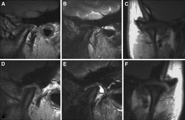
MR images of the TMJ at the initial visit. a–c Right TMJ, d–f left TMJ. a, d Sagittal proton density-weighted images; b, e sagittal T2-weighted images; c, f coronal proton density-weighted images. The sagittal MR images revealed slight erosion of the condyle and joint effusion in the joint space of the TMJ on both sides. The coronal proton density-weighted images clearly revealed synovial proliferation (arrowheads) on both sides (c, f). The disk (arrows) was in a normal position in the right TMJ (a) and showed anterior displacement in the left TMJ (d)
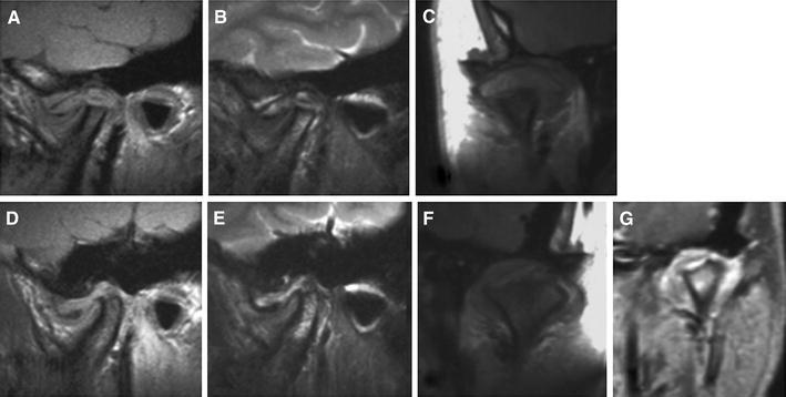
MR images of the TMJ at 1 year after the initial visit. a–c Right TMJ, d–f left TMJ. a, d Sagittal proton density-weighted images; b, e sagittal T2-weighted images; c, f coronal proton density-weighted images of the right TMJ; g post-contrast coronal T1-weighted image of the left TMJ. The sagittal MR images revealed marked deformity of the condyle and articular fossa on both sides. An abnormal signal intensity of the condyle was also noted on the sagittal T2-weighted images (b, e). The coronal proton density-weighted images revealed marked synovial proliferation with bone destruction of the condyle (c, f). On the post-contrast image (g), the synovium and condyle showed strong enhancement
MR images of the TMJ at 2 years after the initial visit. a–c Right TMJ; d–f left TMJ. a, d Sagittal proton density-weighted images; b, e sagittal T2-weighted images; c, f coronal proton density-weighted images. Compared with the findings at 1 year after the initial visit (Fig. 2), the bone destruction of the condyle had progressed on both sides. The synovial proliferation had also become more marked (c, f)
References
-
- Smith HJ, Larheim TA, Aspestrand F. Rheumatic and nonrheumatic disease in the temporomandibular joint: gadolinium-enhanced MR imaging. Radiology. 1992;185:229–234. - PubMed
-
- Sugimoto H, Takeda A, Masuyama J, Furuse M. Early-stage rheumatoid arthritis: diagnostic accuracy of MR imaging. Radiology. 1996;198:185–192. - PubMed
LinkOut - more resources
Full Text Sources