. 2011 Apr;2(2):158-173.

Aging and Cardiac Fibrosis

Affiliations
Free PMC article

Aging and Cardiac Fibrosis

Anna Biernacka et al. Aging Dis. 2011 Apr.
Free PMC article

Abstract

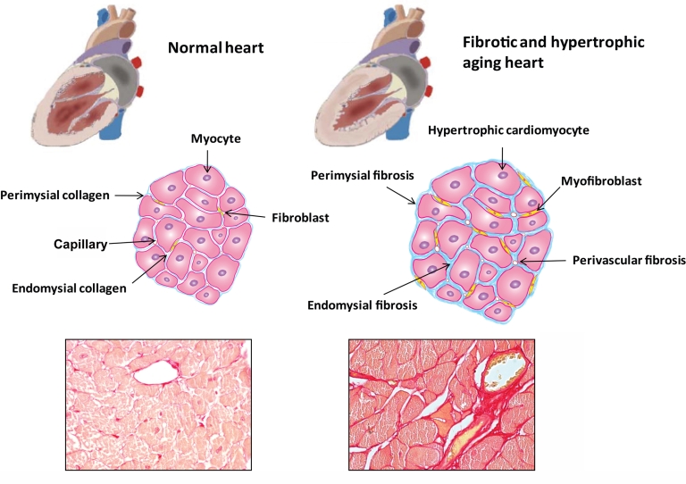
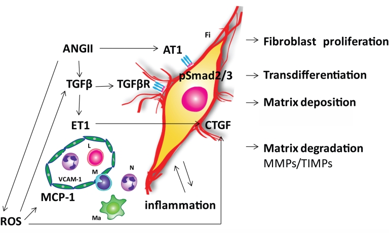
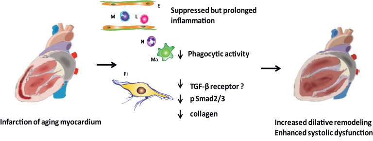
The aging heart is characterized by morphological and structural changes that lead to its functional decline and are associated with diminished ability to meet increased demand. Extensive evidence, derived from both clinical and experimental studies suggests that the aging heart undergoes fibrotic remodeling. Age-dependent accumulation of collagen in the heart leads to progressive increase in ventricular stiffness and impaired diastolic function. Increased mechanical load, due to reduced arterial compliance, and direct senescence-associated fibrogenic actions appear to be implicated in the pathogenesis of cardiac fibrosis in the elderly. Evolving evidence suggests that activation of several distinct molecular pathways may contribute to age-related fibrotic cardiac remodeling. Reactive oxygen species, chemokine-mediated recruitment of mononuclear cells and fibroblast progenitors, transforming growth factor (TGF)-β activation, endothelin-1 and angiotensin II signaling mediate interstitial and perivascular fibrosis in the senescent heart. Reduced collagen degradation may be more important than increased de novo synthesis in the pathogenesis of aging-associated fibrosis. In contrast to the baseline activation of fibrogenic pathways in the senescent heart, aging is associated with an impaired reparative response to cardiac injury and defective activation of reparative fibroblasts in response to growth factors. Because these reparative defects result in defective scar formation, senescent hearts are prone to adverse dilative remodeling following myocardial infarction. Understanding the pathogenesis of interstitial fibrosis in the aging heart and dissecting the mechanisms responsible for age-associated healing defects following cardiac injury are critical in order to design new strategies for prevention of adverse remodeling and heart failure in elderly patients.


Figures

Figure 1.
Figure 1.
Fibrosis of the aging heart. Cardiac aging is associated with significant alterations in cardiac structure and function. Elderly patients often present with left ventricular hypertrophy and diastolic dysfunction while the systolic function is usually preserved. Age-dependent remodeling of the heart is associated with cardiomyocyte hypertrophy and interstitial fibrosis. In the normal heart, thin layers of perimysium and endomysium surround myocardial bundles and individual myocytes, respectively. The walls of the blood vessels also contain adventitial fibroblasts that contribute to the endomysial collagen network. In the senescent heart, there is hypertrophy of cardiomyocytes, transition of fibroblasts to myofibroblasts and accumulation of extracellular matrix proteins in the interstitium. These alterations lead to perivascular, endomysial and perimysial fibrosis. The histopathologic images show fibrotic remodeling of the heart in aging wild type mice. Picrosirius red staining identifies the collagen network in the myocardium of 2 mo and 24 mo C57BL/6 mice. Senescent hearts (S) display markedly increased collagen content compared to young hearts (Y).
Figure 2.
Figure 2.
Pathways involved in the pathogenesis of cardiac fibrosis in the senescent heart. Angiotensin II (ANG II), reactive oxygen species (ROS), transforming growth factor β (TGF-β) and endothelin-1 (ET-1) signaling appear to play an important role in mediating fibrotic remodeling of the aging heart. ANG II exerts its effects directly through the ANG II type 1 receptor (AT1) and indirectly through induction of TGF-β. Smad 2/3 seems to be a common pathway for these two mechanisms. Age-dependent mitochondrial dysfunction is the major source of ROS in the myocardium. ANG II induces NADPH oxidase dependent generation of ROS. ROS activate TGF-β and upregulate its downstream fibrogenic effector connective tissue growth factor (CTGF). ROS, ANG II and ET-1 induce adhesion molecules in the microvascular endothelium and pro-inflammatory mediator expression. Inflammatory cytokines may induce and activate matrix metalloproteinases (MMPs) enhancing matrix degradation. TGF-β/Smad2/3 signaling promotes fibroblast proliferation, phenotypic conversion to myofibroblasts and the production of extracellular matrix components including fibrillar collagen, fibronectin, and proteoglycans. Changes in the extracellular matrix occur in part due to an imbalance of MMPs and their inhibitors (TIMPs). The altered matrix modifies the pro-survival signals that cardiac myocytes receive from their scaffolding environment, leading to cardiomyocyte loss due to apoptosis or necrosis. Symbols: E, endothelial cell; Fi, fibroblast; L, lymphocyte, Ma, macrophage; M, monocyte; N, neutrophil; TGF-βR, TGF-β receptor; VCAM-1, vascular cell adhesion molecule-1; MCP-1, monocyte chemoattractant protein-1
Figure 3.
Figure 3.
Age-related defects in the inflammatory and reparative response lead to enhanced adverse remodeling following myocardial infarction. Although aging is associated with enhanced baseline inflammation and fibrosis, acute infarction results in suppressed, but prolonged, inflammatory reaction, impaired cardiomyocyte phagocytosis, and markedly diminished collagen deposition in the scar. Evolving evidence suggests that the alterations in post-infarction cardiac repair may be due to impaired responsiveness of senescent fibroblasts to growth factors, such as TGF-β. Whether this is due to an aging-related reduction of TGF-β receptor (TGF-βR) expression by fibroblasts, or reflects impaired TGF-β/Smad2/3 signaling in senescent cells, remains unknown. Diminished collagen deposition may lead to a marked reduction in tensile strength of the scar, resulting in accentuated dilation of the infarcted ventricle. Symbols: E, endothelial cell; Fi, fibroblast; L, lymphocyte, Ma, macrophage; M, monocyte; N, neutrophil

References

    1. Lakatta EG, Levy D. Arterial and cardiac aging: major shareholders in cardiovascular disease enterprises: Part II: the aging heart in health: links to heart disease. Circulation. 2003;107:346–54. - PubMed
    1. Kitzman DW, Gardin JM, Gottdiener JS, Arnold A, Boineau R, Aurigemma G, Marino EK, Lyles M, Cushman M, Enright PL. Importance of heart failure with preserved systolic function in patients > or = 65 years of age. CHS Research Group Cardiovascular Health Study. Am J Cardiol. 2001;87:413–9. - PubMed
    1. Vanoverschelde JJ, Essamri B, Vanbutsele R, d’Hondt A, Cosyns JR, Detry JR, Melin JA. Contribution of left ventricular diastolic function to exercise capacity in normal subjects. J Appl Physiol. 1993;74:2225–33. - PubMed
    1. Gagliano N, Arosio B, Santambrogio D, Balestrieri MR, Padoani G, Tagliabue J, Masson S, Vergani C, Annoni G. Age-dependent expression of fibrosis-related genes and collagen deposition in rat kidney cortex. J Gerontol A Biol Sci Med Sci. 2000;55:B365–72. - PubMed
    1. Abrass CK, Adcox MJ, Raugi GJ. Aging-associated changes in renal extracellular matrix. Am J Pathol. 1995;146:742–52. - PMC - PubMed

LinkOut - more resources