. 2011 Sep 20;44(9):666-76.
doi: 10.1021/ar200148z. Epub 2011 Aug 15.

From mechanism to mouse: a tale of two bioorthogonal reactions

Affiliations
Free PMC article

From mechanism to mouse: a tale of two bioorthogonal reactions

Ellen M Sletten et al. Acc Chem Res. .
Free PMC article

Abstract

Bioorthogonal reactions are chemical reactions that neither interact with nor interfere with a biological system. The participating functional groups must be inert to biological moieties, must selectively reactive with each other under biocompatible conditions, and, for in vivo applications, must be nontoxic to cells and organisms. Additionally, it is helpful if one reactive group is small and therefore minimally perturbing of a biomolecule into which it has been introduced either chemically or biosynthetically. Examples from the past decade suggest that a promising strategy for bioorthogonal reaction development begins with an analysis of functional group and reactivity space outside those defined by Nature. Issues such as stability of reactants and products (particularly in water), kinetics, and unwanted side reactivity with biofunctionalities must be addressed, ideally guided by detailed mechanistic studies. Finally, the reaction must be tested in a variety of environments, escalating from aqueous media to biomolecule solutions to cultured cells and, for the most optimized transformations, to live organisms. Work in our laboratory led to the development of two bioorthogonal transformations that exploit the azide as a small, abiotic, and bioinert reaction partner: the Staudinger ligation and strain-promoted azide-alkyne cycloaddition. The Staudinger ligation is based on the classic Staudinger reduction of azides with triarylphosphines first reported in 1919. In the ligation reaction, the intermediate aza-ylide undergoes intramolecular reaction with an ester, forming an amide bond faster than aza-ylide hydrolysis would otherwise occur in water. The Staudinger ligation is highly selective and reliably forms its product in environs as demanding as live mice. However, the Staudinger ligation has some liabilities, such as the propensity of phosphine reagents to undergo air oxidation and the relatively slow kinetics of the reaction. The Staudinger ligation takes advantage of the electrophilicity of the azide; however, the azide can also participate in cycloaddition reactions. In 1961, Wittig and Krebs noted that the strained, cyclic alkyne cyclooctyne reacts violently when combined neat with phenyl azide, forming a triazole product by 1,3-dipolar cycloaddition. This observation stood in stark contrast to the slow kinetics associated with 1,3-dipolar cycloaddition of azides with unstrained, linear alkynes, the conventional Huisgen process. Notably, the reaction of azides with terminal alkynes can be accelerated dramatically by copper catalysis (this highly popular Cu-catalyzed azide-alkyne cycloaddition (CuAAC) is a quintessential "click" reaction). However, the copper catalysts are too cytotoxic for long-term exposure with live cells or organisms. Thus, for applications of bioorthogonal chemistry in living systems, we built upon Wittig and Krebs' observation with the design of cyclooctyne reagents that react rapidly and selectively with biomolecule-associated azides. This strain-promoted azide-alkyne cycloaddition is often referred to as "Cu-free click chemistry". Mechanistic and theoretical studies inspired the design of a series of cyclooctyne compounds bearing fluorine substituents, fused rings, and judiciously situated heteroatoms, with the goals of optimizing azide cycloaddition kinetics, stability, solubility, and pharmacokinetic properties. Cyclooctyne reagents have now been used for labeling azide-modified biomolecules on cultured cells and in live Caenorhabditis elegans, zebrafish, and mice. As this special issue testifies, the field of bioorthogonal chemistry is firmly established as a challenging frontier of reaction methodology and an important new instrument for biological discovery. The above reactions, as well as several newcomers with bioorthogonal attributes, have enabled the high-precision chemical modification of biomolecules in vitro, as well as real-time visualization of molecules and processes in cells and live organisms. The consequence is an impressive body of new knowledge and technology, amassed using a relatively small bioorthogonal reaction compendium. Expansion of this toolkit, an effort that is already well underway, is an important objective for chemists and biologists alike.


Figures

Figure 1
Figure 1
(A) A generic bioorthogonal chemical reaction between X and Y that proceeds in biological systems. (B) A common experimental platform for biomolecule probing using bioorthogonal chemistry. First, a non-native functional group, often called a “chemical reporter”, is installed in a biomolecule of interest. The modified biomolecule is subsequently labeled using a bioorthogonal chemical reaction.
Figure 2
Figure 2
The number of publications containing the word “bioorthogonal” categorized by year of publication. *The 2011 value is projected based on publications from the first half of the year.
Figure 3
Figure 3
A step-by-step guide to developing a bioorthogonal reaction.
Figure 4
Figure 4
The mechanism of the Staudinger reduction (A) and Staudinger ligation (B).
Figure 5
Figure 5
The Staudinger ligation enables selective biomolecule labeling in a variety of environments. (A) A phosphine–biotin (Phos-biotin) probe for detection of azides through the Staudinger ligation. (B,C) Selective labeling of azide-modified glycoproteins in lysates and on live cells. Jurkat cells were treated with (blue bars) or without (green bars) peracetylated-N-azidoactyl mannosamine (Ac4ManNAz), which is metabolized to N-azidoacetyl neuraminic acid and incorporated into glycoproteins. (B) Lysates were treated with Phos-biotin (250 μM) overnight and analyzed by Western blot probing with an anti-biotin–horse radish peroxidase (HRP) antibody. (C) Live cells were treated with Phos-biotin (250 μM) for 1 h, followed by incubation with a fluorescent avidin protein (FITC-avidin) and analyzed by flow cytometry. (D) Mice were injected with (blue bars) or without (green bars) Ac4ManNAz once daily for 7 d. On the eighth day, phosphine conjugated to the FLAG peptide (Phos-FLAG) was injected into the mice. After 3 h, the mice were sacrificed, and their splenocytes were isolated, incubated with a fluorescent anti-FLAG antibody (FITC-anti-FLAG), and analyzed by flow cytometry. Au = arbitrary units.
Figure 6
Figure 6
(A) A FRET-based fluorogenic phosphine for the Staudinger ligation. (B,C) HeLa cells were grown in the presence (B) or absence (C) of Ac4ManNAz. The cells were washed, incubated with 50 μM 10 for 8 h at 37 °C, and imaged. Green = fluorescein. Blue = Hoechst 33342 nuclear stain. Images were originally published in ref (xi). Copyright 2008, WILEY-VHC. (D) A phosphine–luciferin probe for bioluminescence imaging of azides.
Figure 7
Figure 7
(A) The 1,3-dipolar cycloaddition of azides and linear alkynes to form regioisomeric triazole products. (B) The Cu(I)-catalyzed formal azide–alkyne cycloaddition to yield 1,4-triazole products, also known as CuAAC, a paradigm example of “click chemistry”. (C) The strain-promoted cycloaddition of azides and cyclooctynes to give triazole products, also known as Cu-free click chemistry.
Figure 8
Figure 8
Cyclooctyne selectively reacts with azides through a strain-promoted cycloaddition. (A) A cyclooctyne–biotin probe (OCT-biotin). (B) OCT selectively labels an azide-modified form of the recombinant glycoprotein GlyCAM-IgG. Purified GlyCAM-IgG or azido-GlyCAM-IgG was incubated with 0 or 250 μM OCT-biotin overnight at rt. The samples were analyzed by Western blot probing with an anti-biotin antibody conjugated to HRP. An anti-IgG antibody confirmed equal protein loading. Western blot reprinted with permission from ref (xxvii). Copyright 2004 American Chemical Society. (C) OCT labels live cells in an azide-dependent manner. Jurkat cells were grown in the presence (blue bars) or absence (green bars) of Ac4ManNAz. The cells were incubated with OCT-biotin or Phos-biotin (100 μM) for 1 h at rt, followed by treatment with FITC-avidin, and analyzed by flow cytometry.
Figure 9
Figure 9
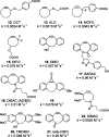
Cyclooctynes synthesized for Cu-free click chemistry in living systems. The second-order rate constants are for the reaction with benzyl azide in acetonitrile (12,(xxvii)13,(xxviii)14,(xxviii)15,(xxix)17,(xxxi)22(xlii)) or methanol (16,(xxxvi)18,(xxxii)19,(xxxiv)20,(xxxv)21(xxxvi)).
Figure 10
Figure 10
The cyclooctynes are superior reagents for labeling azides on cell surfaces. (A,B) Jurkat cells were grown in the presence or absence of Ac4ManNAz. (A) The cells were incubated with Phos-biotin or DIFO-biotin (100 μM) for 1 h at rt, followed by treatment with FITC-avidin, and analyzed by flow cytometry. (B) The cells were treated with BARAC-biotin, DIFO-biotin, or DIBO-biotin (1 μM) for various amounts of time. Each sample was incubated with FITC-avidin and analyzed by flow cytometry. Each point represents the difference between the azide-treated and untreated cells. Au = arbitrary units.
Figure 11
Figure 11
Cyclooctyne–fluorophore conjugates label cells in an azide-dependent manner. CHO (A, B, E–H) or U-2 OS (C, D) cells were grown in the presence (A, C, E, G) or absence (B, D, F, H) of Ac4ManNAz. (A,B) The cells were incubated with DIFO conjugated to Alexa Fluor 488 (DIFO-488, 100 μM) for 1 min at 37 °C, washed, and imaged. (C,D) The cells were incubated with DIBO conjugated to Alexa Fluor 555 (DIBO-555, 30 μM) for 1 h at rt. The cells were then washed, fixed, and imaged. (E,F) The cells were incubated with BARAC conjugated to fluorescein (BARAC-fluorescein, 5 μM) for 5 min, washed, and imaged. (G,H) The cells were incubated with BARAC-fluorescein (250 nM) for 30 min and immediately imaged without washing. Green = DIFO-488 or BARAC-fluorescein; Red = DIBO-555; Blue = Hoechst 33342 nuclear stain.
Figure 12
Figure 12
DIFO–Alexa Fluor conjugates label azides in higher organisms. (A) C. elegans were grown in the presence of Ac4GalNAz and reacted with DIFO-488 (100 μM) followed by DIFO conjugated to Alexa Fluor 568 (DIFO-568, 100 μM) and imaged at their adult stage. Image reprinted with permission from ref (xxxviii). Copyright 2009 American Chemical Society. (B) Zebrafish embryos were metabolically labeled with Ac4GalNAz from 3 to 60 hpf. The fish were sequentially incubated with 100 μM DIFO conjugated to Alexa Fluor 647 (DIFO-647, 60–61 hpf), DIFO-488 (62–63 hpf), and DIFO-555 (72–73 hpf) and imaged by confocal microscopy. During periods in which the zebrafish were not being labeled with DIFO, the fish were bathed in a solution of Ac4GalNAz. Blue = DIFO-647, Green = DIFO-488, Red = DIFO-555. (C) Zebrafish embryos were injected with UDP-GalNAz and a rhodamine–dextran tracer dye. At 7 hpf, the embryos were incubated with DIFO-488 (100 μM) for 1 h and imaged by confocal microscopy. Green = DIFO-488, red = rhodamine–dextran. Image originally published in ref (xl).
Figure 13
Figure 13
The Staudinger ligation is the superior reaction for labeling cell-surface azide-labeled glycoproteins in mice. (A,B) Mice were injected once daily with (blue bars) or without (green bars) Ac4ManNAz for 7 d. On the eighth day (A) Phos-FLAG or DIFO-FLAG or (B) Phos-FLAG or DIMAC-FLAG was injected. After 3 h, the mice were sacrificed, and their splenocytes were isolated, incubated with FITC-anti-FLAG, and analyzed by flow cytometry. Au = arbitrary units.
Figure 14
Figure 14
Cyclooctynes of recent theoretical and experimental interest.

References

    1. Sletten E. M.; Bertozzi C. R. Bioorthogonal Chemistry: Fishing for Selectivity in a Sea of Functionality. Angew. Chem., Int. Ed. 2009, 48, 6974–6998. - PMC - PubMed
    1. Hang H. C.; Yu C.; Kato D. L.; Bertozzi C. R. A Metabolic Labeling Approach towards Proteomic Analysis of Mucin-type O-linked Glycosylation. Proc. Natl. Acad. Sci. U.S.A. 2003, 100, 14846–14851. - PMC - PubMed
    1. Rideout D.; Calogeropoulou T.; Jaworski J.; McCarthy M. Synergism through Direct Covalent Bonding between Agents: A Strategy for Rational Design of Chemotherapeutic Combinations. Biopolymers 1990, 29, 247–262. - PubMed
    1. Griffin B. A.; Adams S. R.; Tsien R. Y. Specific Covalent Labeling of Recombinant Protein Molecules inside Live Cells. Science 1998, 281, 269–272. - PubMed
    1. Saxon E.; Bertozzi C. R. Cell Surface Engineering by a Modified Staudinger Reaction. Science 2000, 287, 2007–2010. - PubMed

Publication types