. 2012 Jan 20;29(2):218-34.
doi: 10.1089/neu.2011.1762. Epub 2011 Sep 29.

Mechanisms of dendritic spine remodeling in a rat model of traumatic brain injury

Affiliations

Mechanisms of dendritic spine remodeling in a rat model of traumatic brain injury

John N Campbell et al. J Neurotrauma. .

Abstract

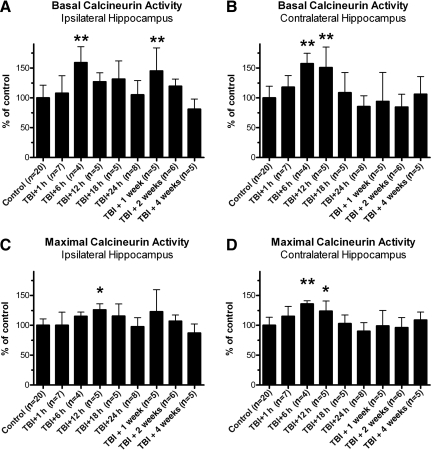
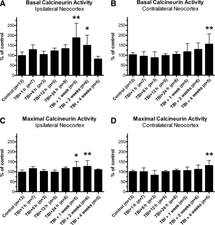
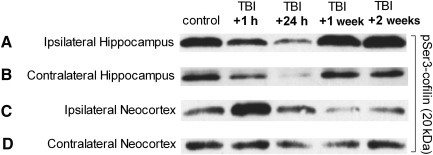
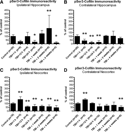
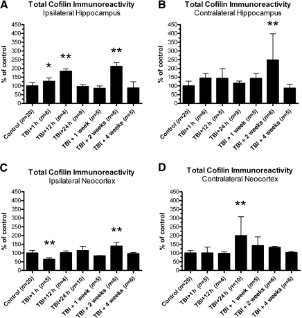
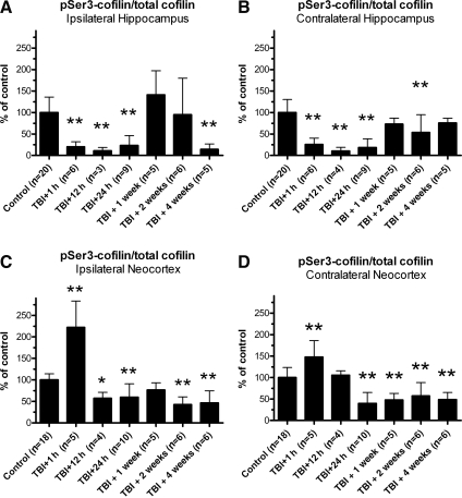
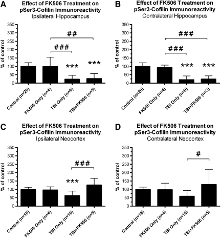
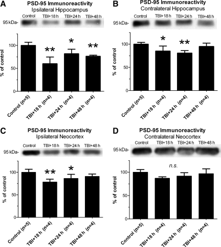
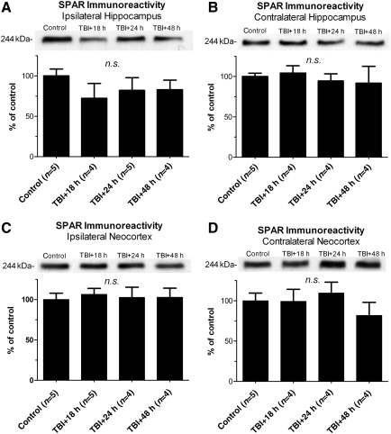
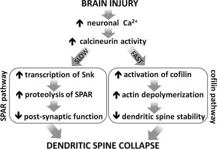
Traumatic brain injury (TBI), a leading cause of death and disability in the United States, causes potentially preventable damage in part through the dysregulation of neural calcium levels. Calcium dysregulation could affect the activity of the calcium-sensitive phosphatase calcineurin (CaN), with serious implications for neural function. The present study used both an in vitro enzymatic assay and Western blot analyses to characterize the effects of lateral fluid percussion injury on CaN activity and CaN-dependent signaling in the rat forebrain. TBI resulted in an acute alteration of CaN phosphatase activity and long-lasting alterations of its downstream effector, cofilin, an actin-depolymerizing protein. These changes occurred bilaterally in the neocortex and hippocampus, appeared to persist for hours after injury, and coincided with synapse degeneration, as suggested by a loss of the excitatory post-synaptic protein PSD-95. Interestingly, the effect of TBI on cofilin in some brain regions was blocked by a single bolus of the CaN inhibitor FK506, given 1 h post-TBI. Overall, these findings suggest a loss of synapse stability in both hemispheres of the laterally-injured brain, and offer evidence for region-specific, CaN-dependent mechanisms.


Figures

FIG. 1.
FIG. 1.
Lateral traumatic brain injury (TBI) enhanced calcineurin phosphatase activity in the hippocampus. Hippocampal tissue was isolated from adult rats at specific time points after lateral fluid percussion injury, homogenized, and subjected to an in vitro assay of calcineurin (CaN) enzymatic activity. CaN activity was measured either in the absence of stimulating cations (basal CaN activity), or in the presence of stimulating cations (maximal CaN activity). In the ipsilateral hippocampus, these experiments revealed increases in basal CaN activity at 6 h and 1 week post-TBI, relative to controls (A). Similarly, in the contralateral hippocampus, increases in basal CaN activity were detected at 6 h and 12 h post-TBI, relative to controls (B). Lateral TBI also affected the maximal activity of CaN, causing increases at 12 h post-TBI in the ipsilateral hippocampus (C), and at 6 h and 12 h post-TBI in the contralateral hippocampus (D), compared to controls. All comparisons were made by one-way analysis of variance with Dunnett's post hoc test (*p<0.05, **p<0.01).
FIG. 2.
FIG. 2.
Lateral traumatic brain injury (TBI) enhanced calcineurin phosphatase activity in the neocortex. Neocortical tissue was isolated from adult rats at specific time points after lateral fluid percussion injury, homogenized, and subjected to an in vitro assay of calcineurin (CaN) enzymatic activity. CaN activity was measured either in the absence (basal CaN activity) or presence (maximal CaN activity) of stimulating cations. In the ipsilateral neocortex, basal CaN activity increased significantly above control levels by 1 week and 2 weeks post-TBI (A). The contralateral neocortex also showed an increase in basal CaN activity, but weeks later, at 4 weeks post-TBI, compared to controls (B). The maximal activity of CaN also changed after lateral TBI, increasing significantly over control levels at 1 week and 2 weeks post-TBI in the ipsilateral neocortex (C), and at 4 weeks post-TBI in the contralateral neocortex (D). All comparisons were made by one-way analysis of variance with Dunnett's post-hoc test (*p<0.05, **p<0.01).
FIG. 3.
FIG. 3.
Lateral traumatic brain injury (TBI) caused apparent changes in serine 3-phosphorylated cofilin (pSer3-cofilin) immunoreactivity. Western blot analysis was performed on crude homogenates of whole ipsilateral and contralateral hemispheres of hippocampus and neocortex dissected from adult rats 1 h, 24 h, 1 week, or 2 weeks after lateral TBI, and from age-matched control rats. The immunoreactivity of pSer3-cofilin was detected with a phospho-specific antibody. Results suggest that the hippocampus and neocortex underwent time- and region-dependent changes in pSer3-immunoreactivity after lateral TBI. In both the ipsilateral and contralateral hippocampi, for example, pSer3-cofilin immunoreactivity appeared to decrease below control levels by 1 h post-TBI, and remained decreased at 24 h post-TBI (A and B). However, by 1 week post-TBI, pSer3-cofilin immunoreactivity appeared to have increased to control levels or beyond. A different trend was observed in the neocortex, where pSer3-cofilin immunoreactivity seemed to initially increase (C), or remain unchanged (D) at 1 h post-TBI, but then decreased at later time points (C and D).
FIG. 4.
FIG. 4.
Lateral traumatic brain injury (TBI) caused time- and region-dependent changes in serine 3-phosphorylated cofilin (pSer3-cofilin) immunoreactivity. The ipsilateral and contralateral hemispheres of the hippocampus and neocortex were dissected from post-TBI rats and their age-matched controls, separately homogenized, and subjected to immunoblotting with antibodies recognizing pSer3-cofilin. The results indicated significant changes in pSer3-cofilin immunoreactivity following lateral TBI, relative to controls. In the ipsilateral hippocampus, for example, pSer3-cofilin decreased significantly at 24 h post-TBI, increased at 2 weeks post-TBI, but then decreased again at 4 weeks post-TBI, relative to controls (A). The contralateral hippocampus also underwent an acute decrease in pSer3-cofilin immunoreactivity at 1 h, 12 h, and 24 h post-TBI, compared to controls (B). The neocortex showed a different pattern of changes in pSer3-cofilin immunoreactivity, with an increase at 1 h post-TBI, followed by decreases at subsequent time points, in both the ipsilateral neocortex (C), and contralateral neocortex (D), relative to controls. Specifically, pSer3-cofilin immunoreactivity decreased below control levels at 12 h post-TBI and all later time points in the ipsilateral neocortex (C), and at 24 h and 4 weeks post-TBI in the contralateral neocortex (D). All comparisons were made by one-way analysis of variance with Dunnett's post-hoc test (*p<0.05, **p<0.01).
FIG. 5.
FIG. 5.
Lateral traumatic brain injury (TBI) generally caused transient increases in total cofilin immunoreactivity. Western blot analyses were repeated using an antibody recognizing cofilin regardless of serine 3 (Ser3) phosphorylation status (total cofilin). Analyses revealed time- and region-dependent changes in total cofilin immunoreactivity, relative to controls. In the ipsilateral hippocampus, for example, total cofilin immunoreactivity was increased at 1 h, 12 h, and 2 weeks post-TBI, relative to controls (A). In the contralateral hippocampus, total cofilin immunoreactivity also increased above control levels, but only weeks later, at 2 weeks post-TBI (B). Unlike all other brain regions tested, the ipsilateral neocortex showed a decrease in total cofilin immunoreactivity at 1 h post-TBI, relative to controls. However, total cofilin immunoreactivity returned to control levels by 12 h post-TBI, and then increased above control levels weeks later, at 2 weeks post-TBI (C). The contralateral neocortex underwent a delayed increase in total cofilin immunoreactivity at 24 h post-TBI, relative to controls (D). All comparisons were made by one-way analysis of variance with Dunnett's post-hoc test (*p<0.05, **p<0.01).
FIG. 6.
FIG. 6.
Lateral traumatic brain injury (TBI) caused time- and region-dependent changes in the proportion of Ser3-phosphorylated cofilin (pSer3-cofilin). To determine whether TBI affected the proportion of Ser3 phosphorylated cofilin, pSer3-cofilin immunoreactivity data were normalized to total cofilin immunoreactivity data for each sample, yielding a ratio of pSer3-cofilin to total cofilin (p/t cofilin). Analysis of these data identified time- and region-dependent changes in p/t cofilin. In the ipsilateral hippocampus, for example, p/t cofilin was significantly below control levels at 1 h, 12 h, and 24 h post-TBI, and again at 4 weeks post-TBI (A). In the contralateral hippocampus, a similar reduction in p/t cofilin was observed at 1 h, 12 h, and 24 h post-TBI, and then at 2 weeks post-TBI, relative to controls (B). The ipsilateral neocortex showed an increase in p/t cofilin at 1 h post-TBI, followed by decreases in p/t cofilin at 12 h, 24 h, 2 weeks, and 4 weeks post-TBI, relative to controls (C). A similar pattern was observed in the contralateral neocortex, where p/t cofilin was significantly above control levels at 1 h post-TBI, but then dropped below control levels at 24 h, 1 week, 2 weeks, and 4 weeks post-TBI (D). All comparisons were made by one-way analysis of variance with Dunnett's post-hoc test (*p<0.05, **p<0.01).
FIG. 7.
FIG. 7.
A 1-h post-traumatic brain injury (TBI) administration of FK-506 prevented the 24 h post-TBI loss of pSer3-cofilin immunoreactivity in the neocortex but not in the hippocampus. Adult rats received either lateral fluid percussion TBI alone (TBI only,), TBI followed by a 1-h post-TBI injection of FK-506 (5 mg/kg IP; TBI+FK-506), FK-506 injection but no TBI (FK-506 only), or no treatment (control). All groups were sacrificed at 24 h post-TBI (or 23 h post-injection), along with equivalently-aged control rats. The ipsilateral and contralateral hemispheres of the hippocampus and neocortex were dissected, separately homogenized, and subjected to immunoblotting with antibodies recognizing the serine 3-phosphorylated cofilin (pSer3-cofilin). Statistical analysis identified region-specific effects of FK-506 treatment on pSer3-cofilin phosphorylation. For example, FK-506 treatment did not prevent the loss of pSer3-cofilin at 24 h post-TBI, in either the ipsilateral hippocampus (A) or contralateral hippocampus (B). In the neocortex, however, FK-506 treatment completely blocked the loss of pSer3-cofilin at 24 h post-TBI in both the ipsilateral (C) and contralateral (D) hemispheres. All comparisons were made by one-way analysis of variance with Tukey's post-hoc test (***p<0.001 versus controls; #p<0.05 versus the other bracketed group; ##p<0.01 versus the other bracketed group; ###p<0.001 versus the other bracketed group).
FIG. 8.
FIG. 8.
Lateral traumatic brain injury (TBI) caused a loss of PSD-95 immunoreactivity in forebrain. The ipsilateral and contralateral hemispheres of the hippocampus and neocortex were dissected from adult rats at 18 h, 24 h, and 48 h after lateral fluid percussion TBI, separately homogenized, and subjected to immunoblotting with antibody recognizing the post-synaptic protein PSD-95. Lateral TBI caused a significant loss of PSD-95 immunoreactivity at 18 h, 24 h, and 48 h post-TBI in the ipsilateral hippocampus (A), and at 18 h and 24 h post-TBI in the contralateral hippocampus (B), relative to controls. In the ipsilateral neocortex, PSD-95 immunoreactivity also decreased significantly below control levels at 18 h and 24 h post-TBI (C). In the contralateral neocortex, however, PSD-95 immunoreactivity did not differ significantly from controls at any time point tested (D). All comparisons were made by one-way analysis of variance with Dunnett's post-hoc test (*p<0.05; **p<0.01; n.s., no significant differences).
FIG. 9.
FIG. 9.
Effect of lateral traumatic brain injury (TBI) on spine-associated Rap guanosine triphosphatase activating protein (SPAR) immunoreactivity. The ipsilateral and contralateral hemispheres of the hippocampus and neocortex were dissected from adult rats at 18 h, 24 h, and 48 h after lateral fluid percussion TBI, separately homogenized, and subjected to immunoblotting with an antibody recognizing SPAR. No change in SPAR immunoreactivity was detected at any time point tested, in the ipsilateral hippocampus (A), contralateral hippocampus (B), ipsilateral neocortex (C), or contralateral neocortex (D), relative to controls. All comparisons were made by one-way analysis of variance with Dunnett's post-hoc test (n.s., no significant differences).
FIG. 10.
FIG. 10.
Calcineurin-dependent mechanisms by which brain injury could cause dendritic spine collapse. Brain injury can cause an increase in the activity of the calcium-sensitive phosphatase calcineurin (CaN). CaN-dependent signaling can then lead to dendritic spine collapse through different signaling pathways. In the neocortex, for example, CaN activity may lead to a rapid de-phosphorylation/activation of the actin-depolymerizing protein cofilin. Excessive cofilin activity could disrupt the spine's actin-rich cytoskeleton, resulting in spine shrinkage or de-stabilization (e.g., Zhou et al., 2004). In the hippocampus, however, a different mechanism may be involved, a mechanism involving CaN but not cofilin. For example, an injury-induced increase in CaN activity can lead to a transcriptional upregulation of serum-induced kinase (Snk), ultimately resulting in the targeted proteolysis of a spine-stabilizing protein, spine-associated Rap guanosine triphosphatase activating protein (SPAR; Pak and Sheng, 2003). SPAR loss is associated with degeneration of post-synaptic function and structure, including spine loss (Pak and Sheng, ; Pak et al., ; Seeburg et al., 2008). Therefore, cofilin activation and SPAR proteolysis represent different mechanisms by which CaN may cause spine loss. Evidence from the present study favors cofilin activation as a regional mechanism of spine loss after TBI, but does not rule out focal involvement of SPAR proteolysis.

Similar articles

Cited by

References

    1. Ansari M.A. Roberts K.N. Scheff S.W. A time course of contusion-induced oxidative stress and synaptic proteins in cortex in a rat model of TBI. J. Neurotrauma. 2008b;25:513–526. - PubMed
    1. Ansari M.A. Roberts K.N. Scheff S.W. Oxidative stress and modification of synaptic proteins in hippocampus after traumatic brain injury. Free Radic. Biol. Med. 2008a;45:443–452. - PMC - PubMed
    1. Bales J.W. Ma X. Yan H.Q. Jenkins L.W. Dixon C.E. Regional calcineurin subunit B isoform expression in rat hippocampus following a traumatic brain injury. Brain Res. 2010;1358:211–220. - PMC - PubMed
    1. Bernstein B.W. Bamburg J.R. ADF/cofilin: a functional node in cell biology. Trends Cell Biol. 2010;20:187–195. - PMC - PubMed
    1. Bradford M.M. A rapid and sensitive method for the quantitation of microgram quantities of protein utilizing the principle of protien-dye binding. Anal. Biochem. 1976;72:248–254. - PubMed

Publication types

LinkOut - more resources