. 2012 Mar;165(5):1424-41.
doi: 10.1111/j.1476-5381.2011.01623.x.

Dynamic monitoring of beating periodicity of stem cell-derived cardiomyocytes as a predictive tool for preclinical safety assessment

Affiliations

Dynamic monitoring of beating periodicity of stem cell-derived cardiomyocytes as a predictive tool for preclinical safety assessment

Yama A Abassi et al. Br J Pharmacol. 2012 Mar.

Abstract

Background and purpose: Cardiac toxicity is a major concern in drug development and it is imperative that clinical candidates are thoroughly tested for adverse effects earlier in the drug discovery process. In this report, we investigate the utility of an impedance-based microelectronic detection system in conjunction with mouse embryonic stem cell-derived cardiomyocytes for assessment of compound risk in the drug discovery process.

Experimental approach: Beating of cardiomyocytes was measured by a recently developed microelectronic-based system using impedance readouts. We used mouse stem cell-derived cardiomyocytes to obtain dose-response profiles for over 60 compounds, including ion channel modulators, chronotropic/ionotropic agents, hERG trafficking inhibitors and drugs known to induce Torsades de Pointes arrhythmias.

Key results: This system sensitively and quantitatively detected effects of modulators of cardiac function, including some compounds missed by electrophysiology. Pro-arrhythmic compounds produced characteristic profiles reflecting arrhythmia, which can be used for identification of other pro-arrhythmic compounds. The time series data can be used to identify compounds that induce arrhythmia by complex mechanisms such as inhibition of hERG channels trafficking. Furthermore, the time resolution allows for assessment of compounds that simultaneously affect both beating and viability of cardiomyocytes.

Conclusions and implications: Microelectronic monitoring of stem cell-derived cardiomyocyte beating provides a high throughput, quantitative and predictive assay system that can be used for assessment of cardiac liability earlier in the drug discovery process. The convergence of stem cell technology with microelectronic monitoring should facilitate cardiac safety assessment.


Figures

Figure 2
Figure 2
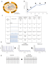
Dynamic monitoring and characterization of mESCC beating using impedance-based detection. (A) Diagram of interdigitated gold microelectronic sensors etched in the bottom of each well of 96 well E-Plate. Application of a low-voltage AC signal generates an electric field between the electrodes which is further impeded by the presence of adherent cardiomyocytes. The interaction of beating cardiomyocyte membranes with the surface of microelectrodes modulates the impedance readout in a cyclical manner. (B) mESCCs were seeded in the wells of the E-Plate and allowed to adhere and form a syncytium. The cells were cultured for up to 96 h and monitored by RTCA Cardio system at regular intervals. The media in the wells were changed once daily. (C) Beating activity and profile of mESCCs recorded by the RTCA Cardio system at indicated time points after cell seeding. The beating rate (1 per min), amplitude (delta CI), beat duration (IBD50; ms), time to max (ms) and decay time (ms) were quantified using the RTCA Cardio software and as described in the Methods section. The data represent the mean of 8 wells ± SD. A total duration of 5 s recording time is displayed. (D) Blebbistatin, an inhibitor of myosin heavy chain ATPase activity, inhibits beating activity of mESCCs, which is restored by washing out the compound and replacing by normal growth media. (E) Blebbistatin treatment of mESCC has no effect on field potential recording as measured by MEAs.
Figure 1
Figure 1
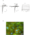
Functional and cell biological characterization of mESCCs. (A) Whole cell currents from mESCC recorded in the voltage clamp mode with the automated patch clamp system Port-a-Patch® from Nanion reveal typical cardiac ion currents: INa, ICa, and IK. (B) Double immunostaing for cardiac α-actinin (green) and Cx43 (red) and nuclei are stained with DAPI (blue). Typical cross striation of cardiac myocytes is shown by staining for cardiac α-actinin. Membrane localization of gap junctions is demonstrated by immunostaining for Cx43.
Figure 3
Figure 3
Pharmacological characterization of mESCC. The cells were seeded in the wells of the E-Plate, monitored for 3 days using the RTCA Cardio system and treated with the indicated concentrations of each compound. The beating activity was recorded by the RTCA Cardio system. For each compound at the indicated time points, 5 s of beating activity is displayed. The beating rate for each interval of beating activity is displayed as beats·min−1± SD; the data shown are one representative recording from a total of at least three separate experiments. (A) Isradipine, an L-type voltage-gated calcium channel inhibitor. (B) (S)-(-)Bay K 8644, an agonist of L-Type voltage-gated calcium channels. (C) Chromanol 293B, inhibitor of the slow delayed rectifier K+ current.
Figure 4
Figure 4
Pharmacological assessment of a ERG channel inhibitor, a sodium channel inhibitor and a chronotropic agent on mESCC beating using the RTCA Cardio system. Dose- and time-dependent effect of (A) E4031, an inhibitor of ERG type K+ channel; 14 s of beating activity is shown (B) TTX, inhibitor of voltage-gated Na+ channel; (C) isoprenaline, a chronotrpic/ionotropic agent and agonist of the β-adrenoceptor; and (D) 0.25% final DMSO concentration. In B, C and D 5 s of beating activity is displayed.
Figure 5
Figure 5
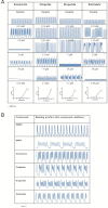
Mechanism-based cardiotoxicity profiling using the RTCA Cardio system. (A) The indicated drugs which have been withdrawn from the market due to association with increased incidence of TdP arrhythmia were screened in a dose-response manner using mESCCs. For each compound, a total of 5 s of beating activity is displayed. For each compound, the time- and dose-dependent response was first analysed and the time points that displayed optimal dose-response relationship were chosen for analysis. For astemizole, cisapride, droperide and sertindole, the dose-response profiles are shown at 30 min, 15 min, 180 min and 165 min after compound addition, respectively. The bottom row shows the dose-response for each of the compounds at the indicated time points based on calculation of beat duration parameter (B) Plateau oscillation profiles are induced by all four compounds tested in A as well as E-4031, indicating a common underlying mechanism; at total of 9 s of beating profile recording is displayed for each of the compounds.
Figure 6
Figure 6
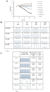
Functional multiplexing using the time resolution of the RTCA Cardio system. mESCCs were seeded in the wells of E-Plates and monitored by the RTCA Cardio system. On the third day, the cells were treated with increasing concentrations of doxorubicin and global cellular responses as well as beating activity were monitored at defined intervals. (A) mESCCs treated with increasing concentrations of doxorubicin. Dose-dependent impedance-based cellular profiles were monitored for up to 24 h after compound treatment (B). A total of 5 s of recording is shown for each dose at the given time point. The beating activity is quantified in terms of beating rate and displayed in each box. (C) mESCCs were seeded in the wells of E-Plate and on day 3 treated with 20 µM pentamidine. The beating activity was monitored at the indicated time windows after compound treatment and quantified based on beat duration. (D) Beating profiles of safe compounds (aspirin, acetaminophen, ibuprofen, clopidogrel and atorvastatin) and compounds with reported cardiac liability (moxifloxacin and quinidine). For each compound, the reported Cmax as well as the concentration tested in the assay is shown. (E) Time-dependent analysis of the compounds in (D) using two analysis parameters; TIBD50 for beat duration and normalized beating rate (BR) for assessment of BR relative to baseline recording immediately before compound addition.
Figure 6
Figure 6
Functional multiplexing using the time resolution of the RTCA Cardio system. mESCCs were seeded in the wells of E-Plates and monitored by the RTCA Cardio system. On the third day, the cells were treated with increasing concentrations of doxorubicin and global cellular responses as well as beating activity were monitored at defined intervals. (A) mESCCs treated with increasing concentrations of doxorubicin. Dose-dependent impedance-based cellular profiles were monitored for up to 24 h after compound treatment (B). A total of 5 s of recording is shown for each dose at the given time point. The beating activity is quantified in terms of beating rate and displayed in each box. (C) mESCCs were seeded in the wells of E-Plate and on day 3 treated with 20 µM pentamidine. The beating activity was monitored at the indicated time windows after compound treatment and quantified based on beat duration. (D) Beating profiles of safe compounds (aspirin, acetaminophen, ibuprofen, clopidogrel and atorvastatin) and compounds with reported cardiac liability (moxifloxacin and quinidine). For each compound, the reported Cmax as well as the concentration tested in the assay is shown. (E) Time-dependent analysis of the compounds in (D) using two analysis parameters; TIBD50 for beat duration and normalized beating rate (BR) for assessment of BR relative to baseline recording immediately before compound addition.

References

    1. Abassi YA, Xi B, Zhang W, Ye P, Kirstein SL, Gaylord MR, et al. Kinetic cell-based morphological screening: prediction of mechanism of compound action and off-target effects. Chem Biol. 2009;16:712–723. - PMC - PubMed
    1. Alexander SPH, Mathie A, Peters JA. Guide to Receptors and Channels (GRAC), 5th edn. Br J Pharmacol. 2011;164(Suppl. 1):S1–S324. - PMC - PubMed
    1. Arrigoni C, Crivori P. Assessment of QT liabilities in drug development. Cell Biol Toxicol. 2007;23:1–13. - PubMed
    1. Atienza JM, Yu N, Kirstein SL, Xi B, Wang X, Xu X, et al. Dynamic and label-free cell-based assays using the real-time cell electronic sensing system. Assay Drug Dev Technol. 2006a;4:597–607. - PubMed
    1. Atienza JM, Yu N, Wang X, Xu X, Abassi Y. Label-free and real-time cell-based kinase assay for screening selective and potent receptor tyrosine kinase inhibitors using microelectronic sensor array. J Biomol Screen. 2006b;11:634–643. - PubMed

MeSH terms