. 2011 Oct;179(4):1719-32.
doi: 10.1016/j.ajpath.2011.06.033. Epub 2011 Aug 11.

Angiotensin II contributes to podocyte injury by increasing TRPC6 expression via an NFAT-mediated positive feedback signaling pathway

Affiliations

Angiotensin II contributes to podocyte injury by increasing TRPC6 expression via an NFAT-mediated positive feedback signaling pathway

Tom Nijenhuis et al. Am J Pathol. 2011 Oct.

Abstract

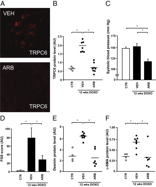
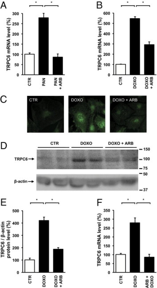
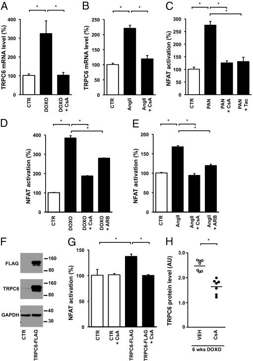
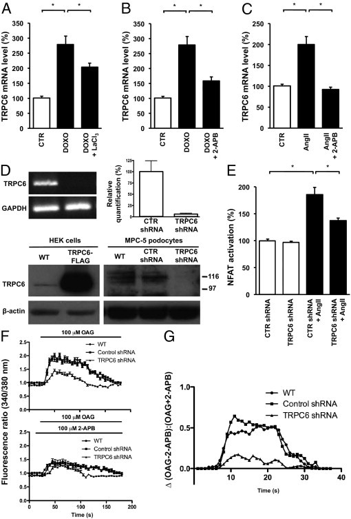
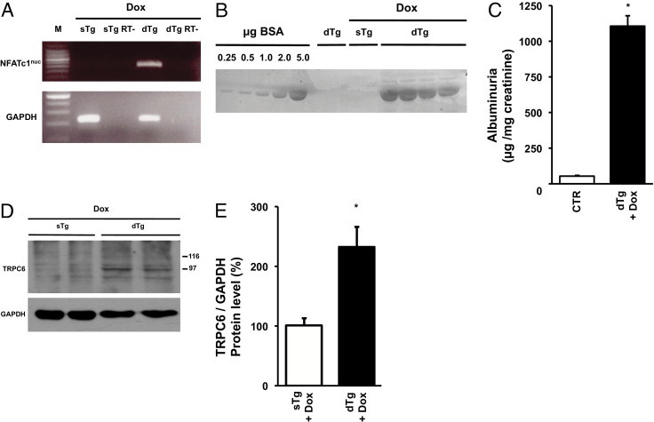
The transient receptor potential channel C6 (TRPC6) is a slit diaphragm-associated protein in podocytes involved in regulating glomerular filter function. Gain-of-function mutations in TRPC6 cause hereditary focal segmental glomerulosclerosis (FSGS), and several human acquired proteinuric diseases show increased glomerular TRPC6 expression. Angiotensin II (AngII) is a key contributor to glomerular disease and may regulate TRPC6 expression in nonrenal cells. We demonstrate that AngII regulates TRPC6 mRNA and protein levels in cultured podocytes and that AngII infusion enhances glomerular TRPC6 expression in vivo. In animal models for human FSGS (doxorubicin nephropathy) and increased renin-angiotensin system activity (Ren2 transgenic rats), glomerular TRPC6 expression was increased in an AngII-dependent manner. TRPC6 expression correlated with glomerular damage markers and glomerulosclerosis. We show that the regulation of TRPC6 expression by AngII and doxorubicin requires TRPC6-mediated Ca(2+) influx and the activation of the Ca(2+)-dependent protein phosphatase calcineurin and its substrate nuclear factor of activated T cells (NFAT). Accordingly, calcineurin inhibition by cyclosporine decreased TRPC6 expression and reduced proteinuria in doxorubicin nephropathy, whereas podocyte-specific inducible expression of a constitutively active NFAT mutant increased TRPC6 expression and induced severe proteinuria. Our findings demonstrate that the deleterious effects of AngII on podocytes and its pathogenic role in glomerular disease involve enhanced TRPC6 expression via a calcineurin/NFAT positive feedback signaling pathway.


Figures

Figure 1
Figure 1
Glomerular TRPC6 expression in the doxorubicin nephropathy (DN) rat model. Representative images of glomerular immunolabeling for TRPC6 in control rats (CTR) and rats with DN (A). TRPC6 expression was determined by semiquantitative analyses of immunofluorescence signals in doxorubicin (DOXO)–exposed and contralateral control (CTR) kidneys, 6, 18, and 30 weeks after treatment (B). Correlation between glomerular TRPC6 expression in the DOXO-exposed kidney and proteinuria in the unilateral DN model (C). Correlation between semiquantitative scoring for TRPC6 protein level and the FGS score in DOXO-exposed (closed dots) and contralateral control (open dots) kidneys (D). TRPC6 protein (E) and mRNA levels (F) in bilateral DN. Co-localization studies for TRPC6 with the marker for injured podocytes desmin (G), the mesangial injury marker α-SMA (H), or HS in the GBM (I). *P < 0.05.
Figure 2
Figure 2
Effect of angiotensin receptor blockade on glomerular TRPC6 expression in bilateral doxorubicin nephropathy. Glomerular TRPC6 expression was determined semiquantitatively in control animals (CTR), vehicle-treated doxorubicin nephropathy rats (VEH), and L158,809-treated doxorubicin nephropathy rats (ARB) (A and B). In addition, blood pressure (C), the FGS score (D), expression of the podocyte injury marker desmin (E), and the mesangial injury marker α-SMA (F) were determined in the respective groups. DOXO, doxorubicin. *P < 0.05.
Figure 3
Figure 3
Effect of angiotensin receptor blockade on TRPC6 mRNA and protein levels in in vitro podocyte injury. TRPC6 mRNA levels were determined in untreated cultured podocytes (CTR) and podocytes incubated for 24 hours with PAN or co-incubated with losartan (PAN + ARB) (n = 5–6 separate podocyte cultures per experimental condition) (A). The effect of doxorubicin (DOXO) and co-incubation with DOXO and losartan (DOXO + ARB) on TRPC6 mRNA levels (B). TRPC6 protein expression visualized by immunocytochemistry of cultured podocytes (C). Immunoblot analysis of cell lysates derived from DOXO- and/or losartan-treated podocytes for TRPC6 and β-actin. Molecular weights indicated in kiloDaltons.(D). Intensity of TRPC6 immunoblot signals was quantified by densitometry and TRPC6 protein levels were normalized to β-actin (E). Effect of preincubation with ARB 1 hour before DOXO application (ARB + DOXO) on TRPC6 mRNA levels (F). *P < 0.05.
Figure 4
Figure 4
Effects of AngII, ARBs, ACEis, and renin on TRPC6 expression in podocytes in vitro and in vivo. Glomerular TRPC6 expression was determined by semiquantitative analyses of immunofluorescence signals in vehicle-treated (VEH) and AngII-treated rats (A). TRPC6 mRNA levels in untreated differentiated cultured podocytes (CTR) and podocytes incubated with AngII (B). Glomerular TRPC6 expression was determined semiquantitatively by immunohistochemistry in Ren2 overexpressing transgenic (Ren2 Tg) rats and wild-type (WT) controls, which were treated with VEH or candesartan (ARB) (C). TRPC6 mRNA levels in CTR, podocytes incubated with doxorubicin (DOXO), and increasing concentrations of renin (REN) and losartan (ARB) (D). Glomerular TRPC6 expression in VEH and lisinopril-treated (ACEi) doxorubicin nephropathy rats (E). Systolic blood pressure (F) and the FGS score (G) in VEH and lisinopril-treated doxorubicin nephropathy rats. TRPC6 mRNA levels in CTR, podocytes incubated with DOXO, and DOXO-treated podocytes in the presence or absence of captopril (ACEi) and/or chymostatin as a blocker of non-ACE, chymase-mediated AngII production (non-ACEi) (H). n = 5 to 6 separate podocyte cultures per experimental condition. *P < 0.05.
Figure 5
Figure 5
Effect of calcineurin inhibition on doxorubicin (DOXO)– and AngII-induced TRPC6 transcription and NFAT activation. TRPC6 mRNA levels in untreated cultured podocytes (CTR) and podocytes incubated with DOXO, as well as podocytes (co)-incubated with DOXO and the calcineurin inhibitor cyclosporine (CsA) (A). The effect of CsA on TRPC6 mRNA levels in AngII-exposed podocytes (B). The induction of NFAT activation in response to PAN was determined using a nuclear NFAT-responsive luciferase reporter construct; thereafter, the effects of the calcineurin inhibitors CsA and tacrolimus (Tac) were studied (C). The effect of DOXO (D) and AngII (E) treatment on NFAT-controlled luciferase expression was determined, as well as after co-treatment with CsA and losartan (ARB). Western blot analysis of total lysates of cultured podocytes infected with a TRPC6-FLAG construct or empty vector (CTR) for FLAG and TRPC6 (F). The GAPDH blot shows equal protein loading. NFAT activation was determined in podocytes infected with a TRPC6-FLAG construct or empty vector in the presence or absence of CsA (G). n = 5 to 6 separate podocyte cultures per experimental condition for all experiments. Glomerular TRPC6 expression was determined in vehicle-treated doxorubicin nephropathy rats (VEH) and CsA-treated doxorubicin nephropathy rats (H). *P < 0.05.
Figure 6
Figure 6
Involvement of Ca2+ influx (through TRPC6 itself) on doxorubicin (DOXO)– and AngII-induced TRPC6 transcription and NFAT activation. TRPC6 mRNA levels in untreated cultured podocytes (CTR) and podocytes incubated with DOXO and DOXO-exposed podocytes co-incubated with LaCl3 (DOXO + LaCl3), which inhibits Ca2+ influx (A). The effect of 2-APB, a TRPC channel blocker, on TRPC6 mRNA levels was evaluated in DOXO- (B) and AngII-treated (C) podocytes. Validation of a podocyte cell line stably transfected with a TRPC6 shRNA construct (D). TRPC6 knockdown was confirmed using semiquantitative RT-PCR, quantitative real-time PCR, and on the protein level Western blot analysis. We determined the effect of AngII on NFAT activation in these podocytes stably expressing a TRPC6 shRNA silencing construct or a control shRNA construct (CTR) (E). n = 3 to 6 separate podocyte cultures per experimental condition for all experiments. *P < 0.05. The OAG-induced Ca2+ response was determined by Fura-2 ratiometry in untransfected podocytes (WT), podocytes stably expressing a TRPC6 shRNA silencing construct, or a CTR shRNA construct, in either the absence or presence of 2-APB (F). The net effect of 2-APB on the OAG-induced Ca2+ response is depicted by calculating the ratio of the change in OAG-induced Ca2+ response in the absence and presence of 2-APB (G).
Figure 7
Figure 7
In vivo effect of NFAT activation in podocytes on TRPC6 expression and proteinuria. RT-PCR analysis of NFATc1nuc transgene expression in glomeruli of podocin-rtTA/tetO-HA-NFATc1nuc mice (A). Double transgenic mice (dTg) were treated with doxycycline (Dox) for 4 days. Single-transgenic rtTA mice (sTg) on Dox served as negative control. Per group glomeruli from three mice were pooled, and half of the RNA sample was not reverse transcribed before PCR and thereby served as no RT control (RT-). Detection of albumin in urine samples from podocin-rtTA/tetO-HA-NFATc1nuc mice by SDS-PAGE and Coomassie staining (B). As controls urine samples from two vehicle-treated dTg mice and two Dox-treated sTg rtTA mice were used. A bovine serum albumin (BSA) gradient (0.25 to 5.0 μg) was run on the same gel to quantify urine albumin signals. Dox-treated dTg mice and control (CTR) mice were analyzed, creating a pool of Dox-treated sTg rtTA mice and vehicle-treated dTg mice (n = 6) as summarized in panel C. TRPC6 and GAPDH protein expression in glomeruli as determined using Western blot analysis (D). As control, Dox-treated sTg rtTA mice were used. Intensity of immunoblot signals was quantified by densitometry and TRPC6 protein levels are depicted relative to GAPDH levels (E). *P < 0.05.
Figure 8
Figure 8
Proposed signaling pathway that regulates the in AngII-dependent TRPC6 expression in podocytes. Results from our study together with previously published data suggest the existence of the following signaling pathways in podocytes mediating AngII-induced TRPC6 expression and podocyte injury. AT1R stimulation by AngII results in Ca2+ influx at least in part mediated by TRPC6. Ca2+-dependent calcineurin activation leads to activation and nuclear translocation of NFAT, which enhances transcription of NFAT-responsive genes such as TRPC6. A consequent increase in TRPC6 protein expression at the cell membrane could result in a positive feedback regulatory circuit, because Ca2+ influx through TRPC6 itself appears to be involved in the signaling pathway leading to increased TRPC6 expression. Our studies show that NFAT activation per se is sufficient to induce TRPC6 transcription and proteinuria. Hypothetically, the proposed feedback mechanism could also result in a persistent calcineurin activation, which was previously demonstrated to lead to the dephosphorylation and degradation of the actin-binding protein synaptopodin (dashed box). The latter was shown to result in cytoskeletal rearrangement and, eventually, proteinuria. Possibly, the latter pathway is involved in the generation of podocyte injury subsequent to AngII-induced TRPC6 expression.

References

    1. Faul C., Asanuma K., Yanagida-Asanuma E., Kim K., Mundel P. Actin up: regulation of podocyte structure and function by components of the actin cytoskeleton. Trends Cell Biol. 2007;17:428–437. - PubMed
    1. Reiser J., Polu K.R., Moller C.C., Kenlan P., Altintas M.M., Wei C., Faul C., Herbert S., Villegas I., Avila-Casado C., McGee M., Sugimoto H., Brown D., Kalluri R., Mundel P., Smith P.L., Clapham D.E., Pollak M.R. TRPC6 is a glomerular slit diaphragm-associated channel required for normal renal function. Nat Genet. 2005;37:739–744. - PMC - PubMed
    1. Winn M.P., Conlon P.J., Lynn K.L., Farrington M.K., Creazzo T., Hawkins A.F., Daskalakis N., Kwan S.Y., Ebersviller S., Burchette J.L., Pericak-Vance M.A., Howell D.N., Vance J.M., Rosenberg P.B. A mutation in the TRPC6 cation channel causes familial focal segmental glomerulosclerosis. Science. 2005;308:1801–1804. - PubMed
    1. Moller C.C., Wei C., Altintas M.M., Li J., Greka A., Ohse T., Pippin J.W., Rastaldi M.P., Wawersik S., Schiavi S., Henger A., Kretzler M., Shankland S.J., Reiser J. Induction of TRPC6 channel in acquired forms of proteinuric kidney disease. J Am Soc Nephrol. 2007;18:29–36. - PubMed
    1. Krall P., Canales C.P., Kairath P., Carmona-Mora P., Molina J., Carpio J.D., Ruiz P., Mezzano S.A., Li J., Wei C., Reiser J., Young J.I., Walz K. Podocyte-specific overexpression of wild type or mutant trpc6 in mice is sufficient to cause glomerular disease. PLoS One. 2010;5:e12859. - PMC - PubMed

Publication types

MeSH terms