

Probing ground and excited states of phospholamban in model and native lipid membranes by magic angle spinning NMR spectroscopy
- PMID: 21839724
- PMCID: PMC3671886
- DOI: 10.1016/j.bbamem.2011.07.040
Probing ground and excited states of phospholamban in model and native lipid membranes by magic angle spinning NMR spectroscopy
Abstract
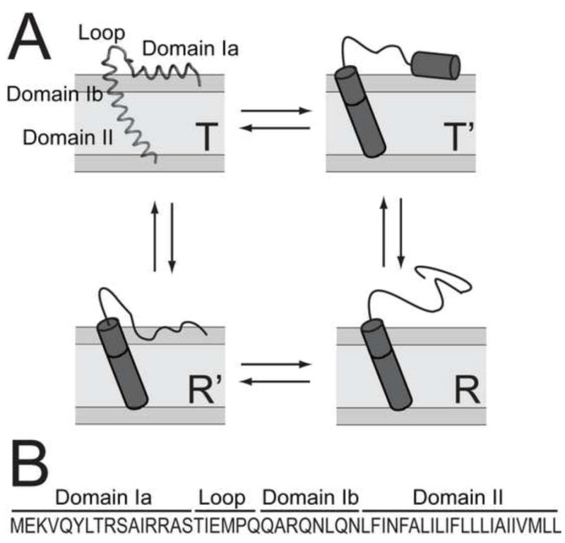
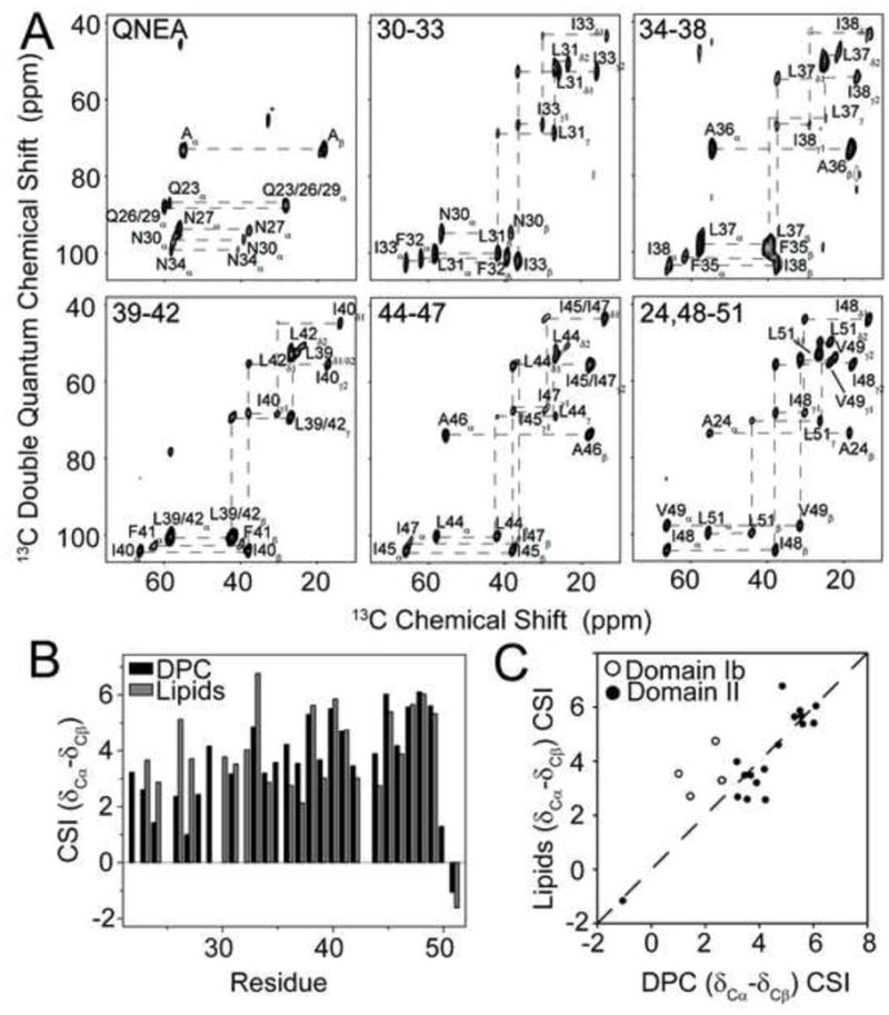
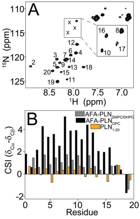
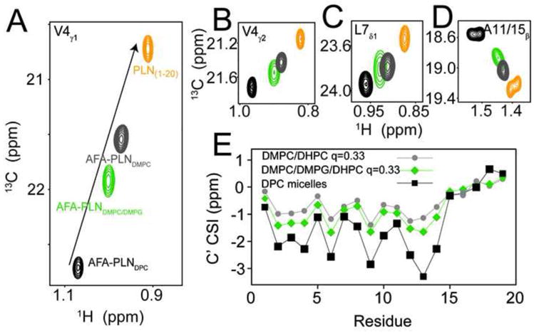
In this paper, we analyzed the ground and excited states of phospholamban (PLN), a membrane protein that regulates sarcoplasmic reticulum calcium ATPase (SERCA), in different membrane mimetic environments. Previously, we proposed that the conformational equilibria of PLN are central to SERCA regulation. Here, we show that these equilibria detected in micelles and bicelles are also present in native sarcoplasmic reticulum lipid membranes as probed by MAS solid-state NMR. Importantly, we found that the kinetics of conformational exchange and the extent of ground and excited states in detergent micelles and lipid bilayers are different, revealing a possible role of the membrane composition on the allosteric regulation of SERCA. Since the extent of excited states is directly correlated to SERCA inhibition, these findings open up the exciting possibility that calcium transport in the heart can be controlled by the lipid bilayer composition. This article is part of a Special Issue entitled: Membrane protein structure and function.
Copyright © 2011 Elsevier B.V. All rights reserved.
Figures
Similar articles
-
Allosteric regulation of SERCA by phosphorylation-mediated conformational shift of phospholamban.Proc Natl Acad Sci U S A. 2013 Oct 22;110(43):17338-43. doi: 10.1073/pnas.1303006110. Epub 2013 Oct 7. Proc Natl Acad Sci U S A. 2013. PMID: 24101520 Free PMC article.
-
Probing excited states and activation energy for the integral membrane protein phospholamban by NMR CPMG relaxation dispersion experiments.Biochim Biophys Acta. 2010 Feb;1798(2):77-81. doi: 10.1016/j.bbamem.2009.09.009. Epub 2009 Sep 23. Biochim Biophys Acta. 2010. PMID: 19781521 Free PMC article.
-
Lipid-mediated folding/unfolding of phospholamban as a regulatory mechanism for the sarcoplasmic reticulum Ca2+-ATPase.J Mol Biol. 2011 May 13;408(4):755-65. doi: 10.1016/j.jmb.2011.03.015. Epub 2011 Mar 17. J Mol Biol. 2011. PMID: 21419777 Free PMC article.
-
The SarcoEndoplasmic Reticulum Calcium ATPase.Subcell Biochem. 2018;87:229-258. doi: 10.1007/978-981-10-7757-9_8. Subcell Biochem. 2018. PMID: 29464562 Review.
-
Flexible P-type ATPases interacting with the membrane.Curr Opin Struct Biol. 2012 Aug;22(4):491-9. doi: 10.1016/j.sbi.2012.05.009. Epub 2012 Jun 28. Curr Opin Struct Biol. 2012. PMID: 22749193 Review.
Cited by
-
Intrinsically disordered HAX-1 regulates Ca2+ cycling by interacting with lipid membranes and the phospholamban cytoplasmic region.Biochim Biophys Acta Biomembr. 2020 Jan 1;1862(1):183034. doi: 10.1016/j.bbamem.2019.183034. Epub 2019 Aug 7. Biochim Biophys Acta Biomembr. 2020. PMID: 31400305 Free PMC article.
-
Secondary structure, backbone dynamics, and structural topology of phospholamban and its phosphorylated and Arg9Cys-mutated forms in phospholipid bilayers utilizing 13C and 15N solid-state NMR spectroscopy.J Phys Chem B. 2014 Feb 27;118(8):2124-33. doi: 10.1021/jp500316s. Epub 2014 Feb 18. J Phys Chem B. 2014. PMID: 24511878 Free PMC article.
-
Structural dynamics and topology of phosphorylated phospholamban homopentamer reveal its role in the regulation of calcium transport.Structure. 2013 Dec 3;21(12):2119-30. doi: 10.1016/j.str.2013.09.008. Epub 2013 Oct 24. Structure. 2013. PMID: 24207128 Free PMC article.
-
The MemMoRF database for recognizing disordered protein regions interacting with cellular membranes.Nucleic Acids Res. 2021 Jan 8;49(D1):D355-D360. doi: 10.1093/nar/gkaa954. Nucleic Acids Res. 2021. PMID: 33119751 Free PMC article.
-
Structures of the excited states of phospholamban and shifts in their populations upon phosphorylation.Biochemistry. 2013 Sep 24;52(38):6684-94. doi: 10.1021/bi400517b. Epub 2013 Sep 11. Biochemistry. 2013. PMID: 23968132 Free PMC article.
References
-
- Farias RN, Bloj B, Morero RD, Sineriz F, Trucco RE. Regulation of allosteric membrane-bound enzymes through changes in membrane lipid compostition. Biochim Biophys Acta. 1975;415:231–251. - PubMed
-
- McDermott A, Polenova T. Solid state NMR: new tools for insight into enzyme function. Curr Opin Struct Biol. 2007;17:617–622. - PubMed
-
- Lange V, Becker-Baldus J, Kunert B, van Rossum BJ, Casagrande F, Engel A, Roske Y, Scheffel FM, Schneider E, Oschkinat H. A MAS NMR study of the bacterial ABC transporter ArtMP. Chembiochem. 2010;11:547–555. - PubMed
Publication types
MeSH terms
Substances
Grants and funding
LinkOut - more resources
Full Text Sources
