. 2011 Aug 16;21(2):366-74.
doi: 10.1016/j.devcel.2011.06.021.

Steep differences in wingless signaling trigger Myc-independent competitive cell interactions

Affiliations

Steep differences in wingless signaling trigger Myc-independent competitive cell interactions

Jean-Paul Vincent et al. Dev Cell. .

Abstract

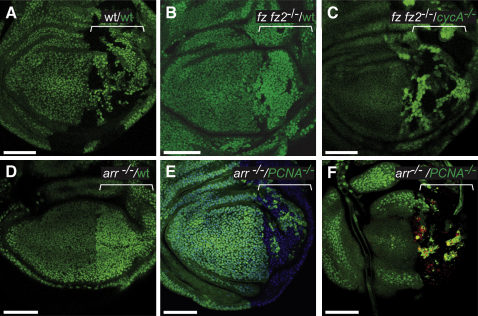
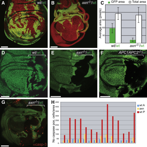
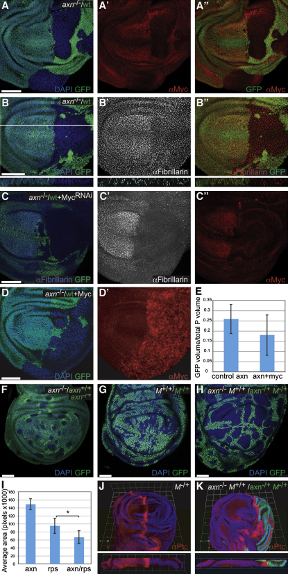
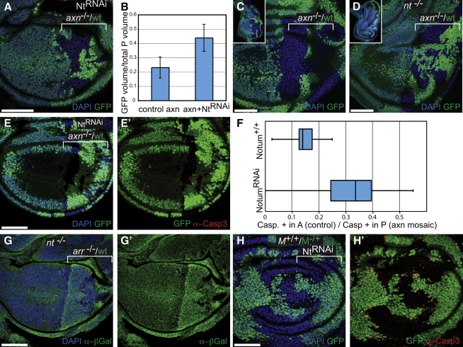
Wnt signaling is a key regulator of development that is often associated with cancer. Wingless, a Drosophila Wnt homolog, has been reported to be a survival factor in wing imaginal discs. However, we found that prospective wing cells survive in the absence of Wingless as long as they are not surrounded by Wingless-responding cells. Moreover, local autonomous overactivation of Wg signaling (as a result of a mutation in APC or axin) leads to the elimination of surrounding normal cells. Therefore, relative differences in Wingless signaling lead to competitive cell interactions. This process does not involve Myc, a well-established cell competition factor. It does, however, require Notum, a conserved secreted feedback inhibitor of Wnt signaling. We suggest that Notum could amplify local differences in Wingless signaling, thus serving as an early trigger of Wg signaling-dependent competition.


Figures

None
Graphical abstract
Figure 1
Figure 1
Survival of Wg-Deficient Cells Depends on the Fitness of Surrounding Cells Twin clones (marked with 2XGFP or the absence of GFP) were generated at maximal frequency in the posterior (P) compartment (marked by a bracket) with engrailed-Gal4, UAS-FLP. On each panel, green lettering describes the GFP-positive and white lettering the GFP-negative tissue. (A) Control disc where both twins are wild-type for Wg signaling (wt). (B) Homozygous fz fz2 mutant cells (GFP-negative) surrounded by wild-type (2XGFP-positive) cells (n = 22/26 discs). (C) fz fz2 (GFP-negative) cells are partially rescued when juxtaposed to growth-compromised cycA mutant (2XGFP-positive) neighbors (n = 11/15 discs). (D–F) Twin clones generated using hedgehog-Gal4, UAS-FLP. (D) Few or no arr mutant (GFP-negative) cells surrounded by wild-type (2XGFP) twin cells survive to late larval stages. (E) arr (GFP-negative) cells are partially rescued when juxtaposed to growth-compromised PCNA mutant (GFP-positive) neighbors (n = 24/24 discs). (F) A basal section from the same disc in (E), processed with antiactivated Caspase- 3 antibody to show apoptotic cells. Most of the Caspase-3 immunoreactivity is in the PCNA−/− territory. All panels show single confocal sections. For all figures scale bars represent 50 μm, Anterior is left and Dorsal on top. Detailed genotypes are listed in the Experimental Procedures.
Figure 2
Figure 2
Cells that Autonomously Overactivate Wg Signaling Outcompete Their Wild-Type Neighbors (A and B) Twin clones induced with escargot-Gal4, UAS-FLP. (A) Wild-type control clones. (B) axin mutant (GFP-negative) and wild-type (GFP-positive) twin clones. (C) Average surface area (±SD) of the pouch and of the GFP-positive domain in 14 control discs (genotype as in A) and 20 experimental discs (as in B). The area of the GFP domain (wild-type) is significantly different in the two conditions (p = 7E-12). (D) Control, wild-type twins generated throughout the P compartment (bracket). (E) As in (B), axin mutant cells affect wild-type (2XGFP) neighbor twins (compare to wild-type 2XGFP cells in control disc in D). (F) APC mutant (GFP-negative) and wild-type (2XGFP) twin clones. (G) Mid-third instar disc of the same genotype as in (E) stained with antiactivated Caspase-3 (red). An optical section through the basal region of the epithelium (where apoptotic cells accumulate) is shown. (H) Quantification of the number of caspase-positive cells per surface area in 14 wing discs of the same genotype as in (G). For each disc, caspase-positive cells were counted throughout the thickness of the wing pouch in three different areas (control A cells in blue, wild-type P cells in red, and axin mutant cells in yellow). Blue and red bars are significantly different p = 3.5E-6). In all panels, micrographs are single sections and GFP is shown in green. DAPI is in red in (A) and (B).
Figure 3
Figure 3
The Competitive Nature of axin Mutant Cells Is Myc Independent and Boosted by Minute-Induced Competition In all the axin−/−/wild-type mosaics, axin mutant cells are GFP-negative and wild-type cells are GFP-positive. (A–A″) Myc expression, as detected with an antibody. (B–B″) Nucleolar size (as judged by anti-Fibrillarin staining), shown here in a mid-third instar disc. Bottom panels show reconstruction of a cross-section at the position indicated by the white line in (B). (C–C″) Uniform downregulation of myc by RNAi throughout the P compartment (with engrailed-Gal4). (C). The efficacy of the RNAi is demonstrated by the downregulation of Fibrillarin (C') and Myc protein levels (C″). (D and D′) Uniform Myc overexpression throughout the P compartment (with engrailed-Gal4, anti-Myc shown in D'). (E) Histogram showing the average fraction of the P compartment occupied by GFP-positive tissue in control discs (genotype as in A or B, n = 8) and in Myc overexpressing discs (genotype as in D, n = 14). Staining and genotypes are indicated on each panel. (F–H) Clones induced simultaneously (using hs-flp) and then allowed to grow for 58 hr before fixation at the end of larval life (see Experimental Procedures). The genotype of GFP-negative cells (“winners”) is indicated in white. (I) Histogram showing the average size (±SD) of the GFP-positive area within the pouch in discs of the same genotype as in (F) (left; n = 4), (G) (middle; n = 8), or (H) (right; n = 4). Asterisk indicates statistical significance; p = 0.027. (J and K) 3D reconstruction of wing discs stained with anti-Ptc (red) to mark the A cells that line the A/P boundary (nonboundary A cells express Ptc weakly). Bottom panels show a cross-section (apical facing up). (J) shows a control disc (Minute−/+) while K shows a wing disc with the A compartment entirely made of GFP-negative, dual competitor cells (genotype as indicated). Note the shift of the Ptc stripe and the reduced size of the P compartment (axin−/+ Minute−/+; GFP-positive). In all panels, GFP is shown in green and DAPI in blue. (C), (D), and (F)–(H) show single sections while (A)–(A ″), (B–B″), (C′ and C″) and (D′) show projections.
Figure 4
Figure 4
notum Mediates Wnt-Induced but Not Minute Competition (A-E) Twin patches of axin mutant (GFP-negative) and axin+ (GFP-positive) cells. (A) notum knockdown throughout the P compartment (with engrailed-Gal4). (B) The average fractional volume (±SD) of the P compartment occupied by GFP-positive tissue in control discs (axin−/−/wild-type mosaics; n = 6; as in Figure 2E) is less than in discs that, in addition, express the notum RNAi transgene (n = 8; genotype as in A); p = 0.00058. (C and D) In notum mutant larvae (D, genotype wf 141/Df3(3L)st-f13), axin+ cells (GFP-positive) are no longer outcompeted by axin mutant cells (n = 19/25; compare to competition in a notum+/+ background as in (C), where competition was observed in 15/18 cases). Top left insets show the imaginal discs in their entirety. (E and E′) Experiments as in (A), stained for activated Caspase-3. (F) For individual discs, the surface density of caspase-positive cells in A (control) and P (outcompeted) cells was measured to generate an A/P ratio (the lower the ratio the stronger cell competition). Such ratios were compared for discs with normal (notum+/+; 14 discs) or knocked down notum activity (notum RNAi; 15 discs). (G and G′) Twin clones of arrow mutant (β-Gal-negative) and wild-type (β-Gal-positive) cells in a notum mutant background (compare with Figures 1D and 1E). (H and H′) Knockdown of notum by RNAi throughout the P compartment with engrailed-Gal4 does not affect Minute−/+ (GFP-positive) versus wild-type (GFP-negative) cell competition. Active Caspase-3 staining in red.

References

    1. Akiyama T., Kamimura K., Firkus C., Takeo S., Shimmi O., Nakato H. Dally regulates Dpp morphogen gradient formation by stabilizing Dpp on the cell surface. Dev. Biol. 2008;313:408–419. - PMC - PubMed
    1. Akong K., Grevengoed E.E., Price M.H., McCartney B.M., Hayden M.A., DeNofrio J.C., Peifer M. Drosophila APC2 and APC1 play overlapping roles in wingless signaling in the embryo and imaginal discs. Dev. Biol. 2002;250:91–100. - PubMed
    1. Ayers K.L., Gallet A., Staccini-Lavenant L., Thérond P.P. The long-range activity of Hedgehog is regulated in the apical extracellular space by the glypican Dally and the hydrolase Notum. Dev. Cell. 2010;18:605–620. - PubMed
    1. Baena-Lopez L.A., Franch-Marro X., Vincent J.P. Wingless promotes proliferative growth in a gradient-independent manner. Sci. Signal. 2009;2 ra60. - PMC - PubMed
    1. Baker N.E., Li W. Cell competition and its possible relation to cancer. Cancer Res. 2008;68:5505–5507. - PubMed

Publication types

MeSH terms