Review
. 2011 Sep;34(9):452-63.
doi: 10.1016/j.tins.2011.07.005. Epub 2011 Aug 15.

Synaptic plasticity in sleep: learning, homeostasis and disease

Affiliations
Review

Synaptic plasticity in sleep: learning, homeostasis and disease

Gordon Wang et al. Trends Neurosci. 2011 Sep.

Abstract

Sleep is a fundamental and evolutionarily conserved aspect of animal life. Recent studies have shed light on the role of sleep in synaptic plasticity. Demonstrations of memory replay and synapse homeostasis suggest that one essential role of sleep is in the consolidation and optimization of synaptic circuits to retain salient memory traces despite the noise of daily experience. Here, we review this recent evidence and suggest that sleep creates a heightened state of plasticity, which may be essential for this optimization. Furthermore, we discuss how sleep deficits seen in diseases such as Alzheimer's disease and autism spectrum disorders might not just reflect underlying circuit malfunction, but could also play a direct role in the progression of those disorders.


Figures

Figure 1
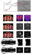
Figure 1. Summary of recent data in support of the Synaptic Homeostasis Hypothesis (SHH)
(a). Synapses, like learning and memories, are known to be affected by circadian rhythms and homeostatic regulation. The SHH posits that synapse accumulation during the day drives a need for synaptic downscaling, which preferentially occurs during sleep. (b-f) Recent studies from diurnal Drosophila melanogaster and Danio rerio have demonstrated increased synapse components or synapse numbers following wake, sleep, or sleep deprivation. Images are not all to the same scale. (b) Bruchpilot (BRP; an essential constituent of the active zone of all synapses) levels were measured in antennal lobes (AL), β lobes of the mushroom bodies (MB), and ellipsoid body of the central complex (CC) in Drosophila [81]. BRP immunofluorescence was found to be increased in animals sleep-deprived for 16 hrs compared to rested controls, shown false-colored on a quantitative scale, with yellow indicating highest levels. (c) Following social enrichment, sleep deprivation was found to lead to the retention of more synaptic terminals in Drosophila olfactory lobes (OL). Discs-large (DLG), a postsynaptic protein, was fused to GFP expressed in PDF neurons via a GAL4;UAS approach (i.e. pdf-GAL4/+::UAS-dlgWT-gfp/+). Social enrichment led to increased numbers of GFP-positive terminals that recovered to baseline levels following sleep but not following sleep deprivation [83]. (d) Synaptotagmin (a presynaptic protein) was fused to EGFP and expressed in the gamma lobe of the MB. Right panels show a higher magnification of the area indicated by the yellow square in the left panel. Sleep-deprived flies were found to contain larger GFP-positive puncta in the MB compared to sleeping controls [82]. Scale bar = 10 μm. (e) Sleep deprivation also interferes with homeostatic downscaling of synapse number in larval zebrafish (7 day old) [84]. Live transgenic fish expressing synaptophysin fused to EGFP in hypocretin neurons (i.e. HCRT:SYPEGFP) displayed significantly more EGFP puncta in axons projecting to the pineal gland (PG) during diurnal wakefulness compared to the nocturnal sleep period. Red arrows depict additional synapses that were not observed during the sleep period in the same fish.(f) Live larval zebrafish expressing HCRT:SYP-EGFP also have more EGFP puncta in hindbrain (HB) projections from the hypocretin neurons [84]. Red arrows indicate additional synapses that were not observed during the sleep period in the same fish. Abbreviations: HB, hindbrain; Hyp, hypothalamus; OT, optic tectum; PG, pineal gland. Reproduced, with permission, from [81] (b), [83] (c), [82] (d).
Figure I
Figure I
Representative images to illustrate the type of images obtained using the different imaging modalities. Top panel: 2-Photon image of transgenic zebrafish expressing EGFP in all HCRT neurons. Middle panel: Array tomography reconstruction of mouse cortical dendrite labeled with yellow fluorescent protein (YFP; green) and synaptotagmin (red) to label synapses. Bottom panel: Segmented microtubule bundles in mouse cortical white matter as imaged using STORM. The colors are pseudocolors to separate out each individual microtubule Image Credits: Gordon Wang (top and middle panels), Nicholas C. Weiler and Xiaowei Zhuang (bottom panel). Abbreviations: not available (N/A).

References

    1. Aserinsky E, Kleitman N. Regularly occurring periods of eye motility, and concomitant phenomena, during sleep. Science. 1953;118:273–274. - PubMed
    1. Jouvet M, et al. On a stage of rapid cerebral electrical activity in the course of physiological sleep. C R Seances Soc Biol Fil. 1959;153:1024–1028. - PubMed
    1. Carskadon MA, Dement WC. Normal Human Sleep: An Overview. Principles and Practice of Sleep Medicine. 2005:13–23.
    1. Tobler I. Effect of forced locomotion on the rest-activity cycle of the cockroach. Behav Brain Res. 1983;8:351–360. - PubMed
    1. Campbell SS, Tobler I. Animal sleep: a review of sleep duration across phylogeny. Neurosci Biobehav Rev. 1984;8:269–300. - PubMed

Publication types