

A full range of mouse sinoatrial node AP firing rates requires protein kinase A-dependent calcium signaling
- PMID: 21840316
- PMCID: PMC3184386
- DOI: 10.1016/j.yjmcc.2011.07.028
A full range of mouse sinoatrial node AP firing rates requires protein kinase A-dependent calcium signaling
Abstract
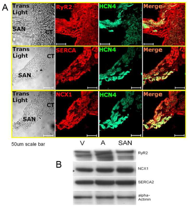
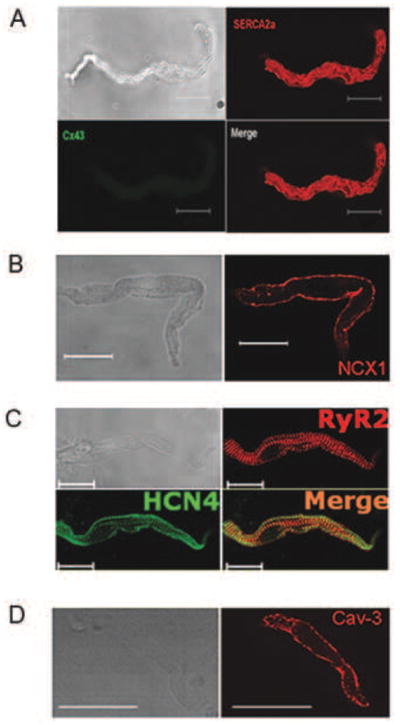
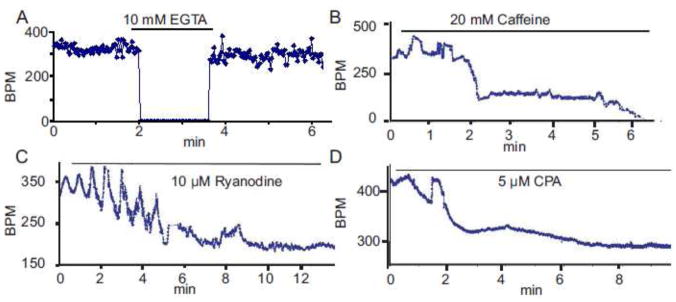
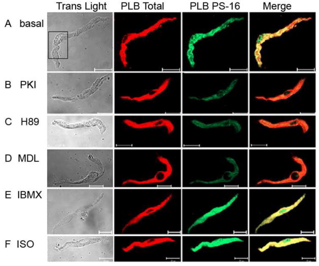
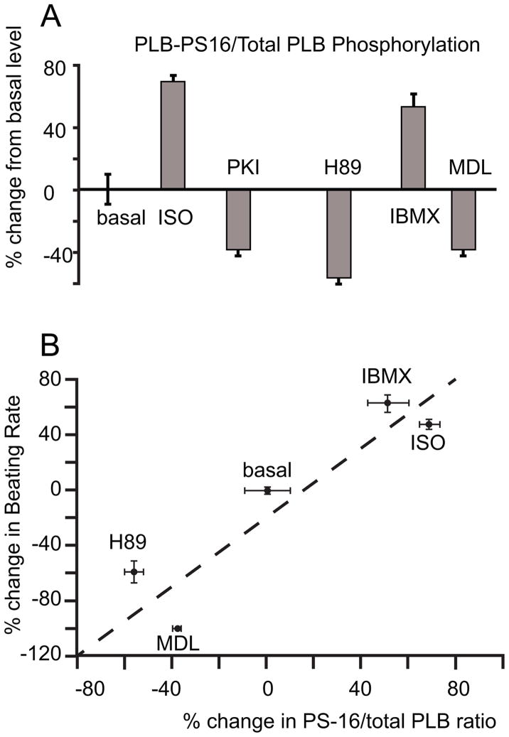
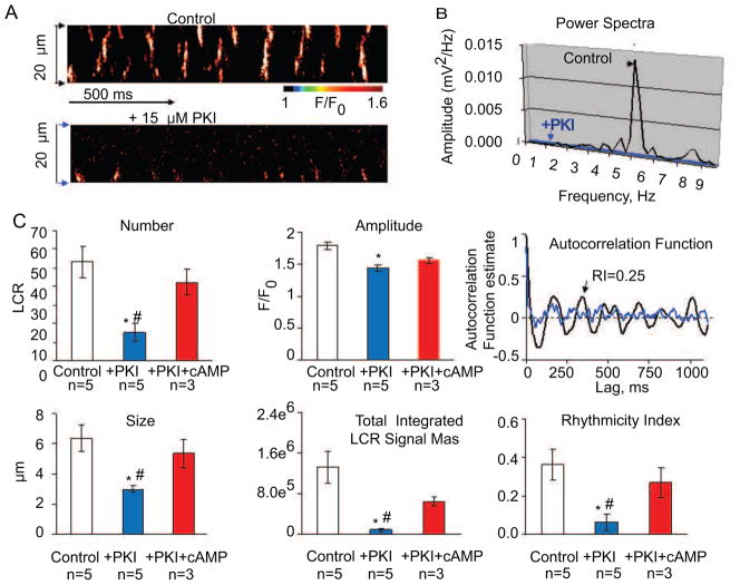
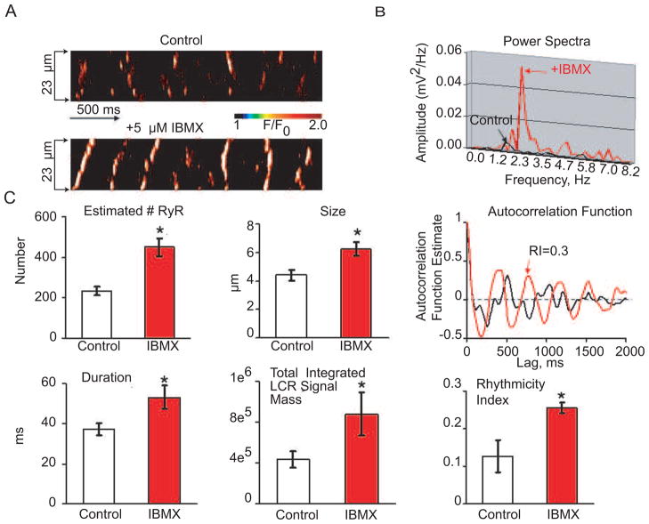
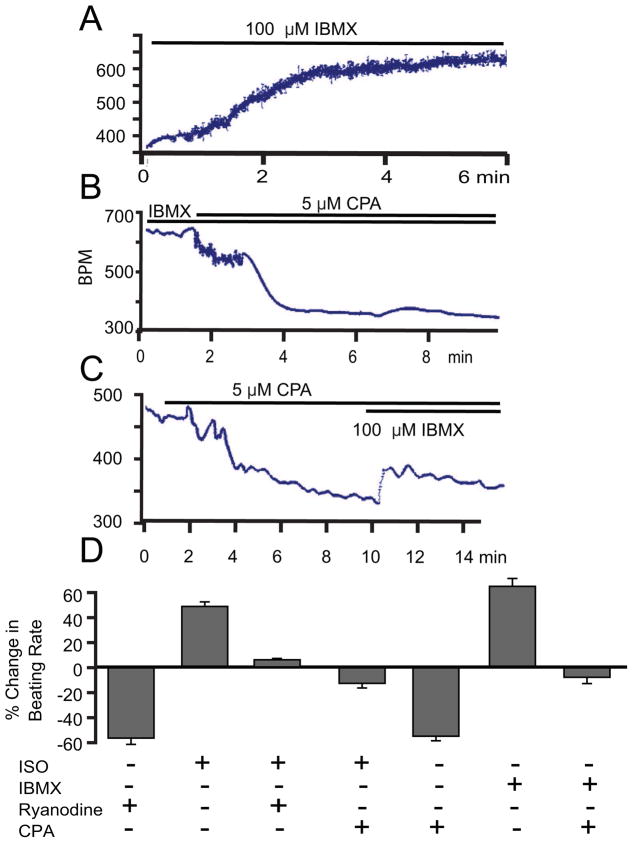
Recent perspectives on sinoatrial nodal cell (SANC)(*) function indicate that spontaneous sarcoplasmic reticulum (SR) Ca(2+) cycling, i.e. an intracellular "Ca(2+) clock," driven by cAMP-mediated, PKA-dependent phosphorylation, interacts with an ensemble of surface membrane electrogenic molecules ("surface membrane clock") to drive SANC normal automaticity. The role of AC-cAMP-PKA-Ca(2+) signaling cascade in mouse, the species most often utilized for genetic manipulations, however, has not been systematically tested. Here we show that Ca(2+) cycling proteins (e.g. RyR2, NCX1, and SERCA2) are abundantly expressed in mouse SAN and that spontaneous, rhythmic SR generated local Ca(2+) releases (LCRs) occur in skinned mouse SANC, clamped at constant physiologic [Ca(2+)]. Mouse SANC also exhibits a high basal level of phospholamban (PLB) phosphorylation at the PKA-dependent site, Serine16. Inhibition of intrinsic PKA activity or inhibition of PDE in SANC, respectively: reduces or increases PLB phosphorylation, and markedly prolongs or reduces the LCR period; and markedly reduces or accelerates SAN spontaneous firing rate. Additionally, the increase in AP firing rate by PKA-dependent phosphorylation by β-adrenergic receptor (β-AR) stimulation requires normal intracellular Ca(2+) cycling, because the β-AR chronotropic effect is markedly blunted when SR Ca(2+) cycling is disrupted. Thus, AC-cAMP-PKA-Ca(2+) signaling cascade is a major mechanism of normal automaticity in mouse SANC.
Published by Elsevier Ltd.
Figures
References
-
- Vinogradova TM, Lyashkov AE, Zhu W, Ruknudin AM, Sirenko S, Yang D, et al. High Basal Protein Kinase A-Dependent Phosphorylation Drives Rhythmic Internal Ca2+ Store Oscillations and Spontaneous Beating of Cardiac Pacemaker Cells. Circ Res. 2006;98:505–14. - PubMed
-
- Vinogradova TM, Sirenko S, Lyashkov AE, Younes A, Li Y, Zhu W, et al. Constitutive phosphodiesterase activity restricts spontaneous beating rate of cardiac pacemaker cells by suppressing local Ca2+ releases. Circ Res. 2008;102:761–9. - PubMed
-
- Ludwig A, Herrmann S, Hoesl E, Stieber J. Mouse models for studying pacemaker channel function and sinus node arrhythmia. Prog Biophys Mol Biol. 2010;98:179–85. - PubMed
Publication types
MeSH terms
Substances
Grants and funding
LinkOut - more resources
Full Text Sources
Other Literature Sources
Research Materials
Miscellaneous
