. 2011 Sep 30;412(4):674-87.
doi: 10.1016/j.jmb.2011.08.004. Epub 2011 Aug 5.

Duplex unwinding and ATPase activities of the DEAD-box helicase eIF4A are coupled by eIF4G and eIF4B

Affiliations

Duplex unwinding and ATPase activities of the DEAD-box helicase eIF4A are coupled by eIF4G and eIF4B

Ali R Özeş et al. J Mol Biol. .

Abstract

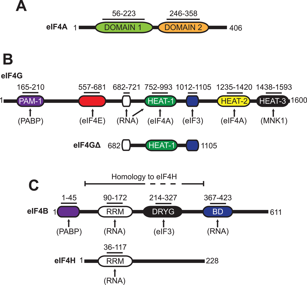
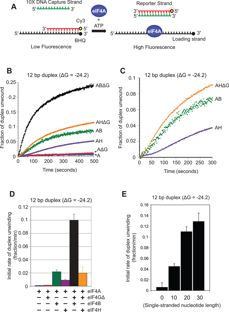
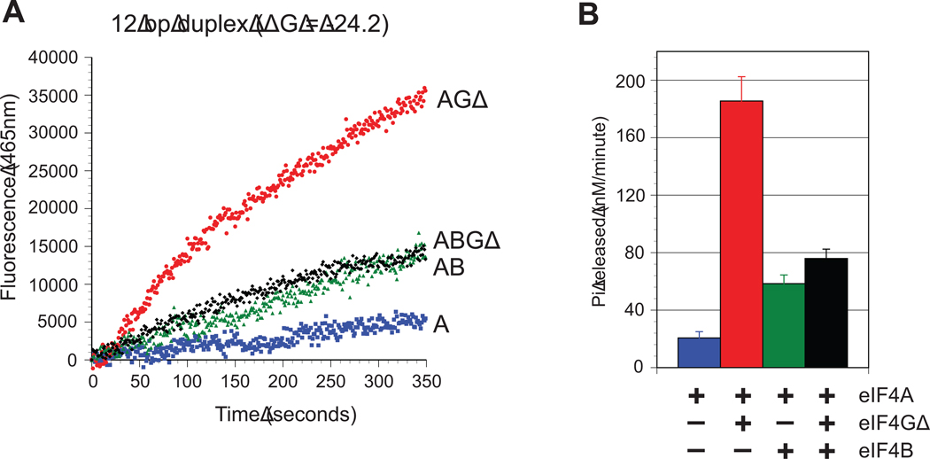
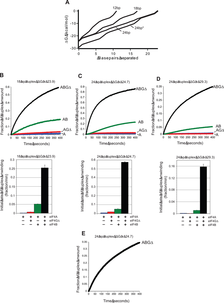
Eukaryotic initiation factor (eIF) 4A is a DEAD-box helicase that stimulates translation initiation by unwinding mRNA secondary structure. The accessory proteins eIF4G, eIF4B, and eIF4H enhance the duplex unwinding activity of eIF4A, but the extent to which they modulate eIF4A activity is poorly understood. Here, we use real-time fluorescence assays to determine the kinetic parameters of duplex unwinding and ATP hydrolysis by these initiation factors. To ensure efficient duplex unwinding, eIF4B and eIF4G cooperatively activate the duplex unwinding activity of eIF4A. Our data reveal that eIF4H is much less efficient at stimulating eIF4A unwinding activity than eIF4B, implying that eIF4H is not able to completely substitute for eIF4B in duplex unwinding. By monitoring unwinding and ATPase assays under identical conditions, we demonstrate that eIF4B couples the ATP hydrolysis cycle of eIF4A with strand separation, thereby minimizing nonproductive unwinding events. Using duplex substrates with altered GC contents but similar predicted thermal stabilities, we further show that the rate of formation of productive unwinding complexes is strongly influenced by the local stability per base pair, in addition to the stability of the entire duplex. This finding explains how a change in the GC content of a hairpin is able to influence translation initiation while maintaining the overall predicted thermal stability.


Figures

Fig. 1
Fig. 1. Schematic representation of eIF4A, eIF4G, eIF4B and eIF4H
Organization of protein domains found in human eIF4A (A), eIF4G (B), eIF4B and eIF4H (C). The eIF4G truncation construct used in this study (eIF4GΔ) is also depicted (B). Functional domains in each protein are shown with their corresponding amino acid regions. Known protein and RNA interaction sites are indicated by arrows. Abbreviations: PAM-1, PABP-binding motif; RRM, RNA recognition motif; DRYG, aspartic acid, arginine, tyrosine and glycine domain; BD, RNA binding domain.
Fig. 2
Fig. 2. Unwinding of an RNA duplex by eIF4A and its accessory proteins
(A) Cartoon depiction of the fluorescent duplex unwinding assay. A reporter RNA strand is modified on its 5′ end with cyanine 3 (Cy3) and annealed to a complimentary loading strand that is modified on its 3′ end with a spectrally paired black hole quencher (BHQ). The loading strand also possesses a 20nt 5′ extension. Addition of eIF4A and ATP to the reaction results in strand separation, which is visualized as an increase in Cy3 fluorescence. Reannealing of the strands is reduced by the presence of a 10-fold molar excess of a DNA capture strand that is complementary to the reporter strand. (B) Representative unwinding time courses of a 12bp duplex substrate (duplex 1; see Materials and Methods) by eIF4A in the absence (blue line) or presence of eIF4GΔ (red line), eIF4H (purple line), eIF4B (green line), eIF4H/4GΔ (orange line) and eIF4B/4GΔ (black line). The zero time point represents the reaction before ATP addition. The change in total fluorescence is converted to the fraction of duplex unwound over time, as described in Materials and Methods. (C) A magnified view of a portion of the unwinding time course from panel B to show the sigmoidal traces for eIF4A in the presence of eIF4H (purple line), eIF4B (green line) and eIF4H/4GΔ (orange line). (D) Initial rates during the initial linear portion of the unwinding time course are determined by linear fits. The initial rate of duplex unwinding by each protein combination is shown as indicated. Error bars represent the standard error from at least three separate experiments. (E) Initial rates of unwinding for a 12bp duplex (duplex 1) possessing either 0, 10, 20 and 30 single-stranded nucleotides added to the 5′ end of the loading strand (see Materials and Methods for sequences). Each assay is carried out in the presence of 50 nM RNA substrate and 1 µM eIF4A/4GΔ/4B. The change in total fluorescence is converted to the fraction of duplex unwound and the initial rate of unwinding during the linear portion of the unwinding time course is shown. Error bars represent the standard error from at least three separate unwinding reactions.
Fig. 3
Fig. 3. ATP hydrolysis by eIF4A and its accessory proteins
(A) Representative time courses of phosphate release by eIF4A on a 12bp duplex (duplex 1) in the absence (blue line) or presence of eIF4GΔ (red line), eIF4B (green line) and eIF4B/4GΔ (black line). The change in RNA-dependent total fluorescence intensity (arbitrary units) of PBP-MDCC at 465 nm against time is shown for each protein combination. The zero time point represents the reaction before ATP addition. (B) Quantitation of the initial rate of RNA-dependent Pi release by eIF4A in the absence or presence of eIF4GΔ, eIF4B and eIF4B/4GΔ, as indicated. The initial change in total fluorescence is converted to the concentration of Pi released (nM min−1). Error bars represent the standard error from at least three separate experiments.
Fig. 4
Fig. 4. Effect of energy landscape on duplex unwinding by eIF4A
(A) The energy landscape of each duplex substrate used is plotted as the change in Gibbs free energy (kcal/mol) per base pair of the duplex unwound. Each trace starts with the 3′ most base pair of the reporter strand and is labeled according to the duplex length. The 24bp* duplex represents the more stable 24bp duplex, as described in the Materials and Methods. (B–D) Upper panels show representative unwinding time courses for duplex substrates possessing different stabilities by eIF4A in the absence (blue line) or presence of eIF4GΔ (red line), eIF4B (green line) and eIF4B/4GΔ (black line). For each time course, the zero time point represents the reaction before ATP addition. The change in total fluorescence is converted to the fraction of duplex unwound over time, as described in Materials and Methods. Unwinding reactions are presented for each protein combination for an 18bp duplex substrate (duplex 2; B), a 24bp duplex (duplex 3, C), and a more stable 24bp duplex (duplex 4; D). Each duplex possesses identical 20 nucleotide 5′ extensions to the loading strand, as described in Materials and Methods. Lower panels in each case show the quantitation of the initial rate of duplex unwinding by eIF4A in the absence (blue bar) or presence of eIF4GΔ (red bar), eIF4B (green bar) and eIF4B/4GΔ (black bar). Initial rates from unwinding reactions during the initial linear portion of each unwinding time course are shown for the 18bp (duplex 2; A), 24bp (duplex 3; B) and more stable 24bp (duplex 4; C) duplexes. Each data set is from at least three separate experiments and error bars represent the standard error. (E) A representative unwinding reaction by an eIF4A/4GΔ/4B complex for a blunt-ended 24bp duplex is shown (duplex 5).

Similar articles

Cited by

References

    1. Rozen F, Edery I, Meerovitch K, Dever TE, Merrick WC, Sonenberg N. Bidirectional RNA helicase activity of eucaryotic translation initiation factors 4A and 4F. Mol Cell Biol. 1990;10:1134–1144. - PMC - PubMed
    1. Rogers GW, Jr, Richter NJ, Merrick WC. Biochemical and kinetic characterization of the RNA helicase activity of eukaryotic initiation factor 4A. J Biol Chem. 1999;274:12236–12244. - PubMed
    1. Rogers GW, Jr, Lima WF, Merrick WC. Further characterization of the helicase activity of eIF4A. Substrate specificity. J Biol Chem. 2001;276:12598–12608. - PubMed
    1. Caruthers JM, Johnson ER, McKay DB. Crystal structure of yeast initiation factor 4A, a DEAD-box RNA helicase. Proc Natl Acad Sci U S A. 2000;97:13080–13085. - PMC - PubMed
    1. Schutz P, Bumann M, Oberholzer AE, Bieniossek C, Trachsel H, Altmann M, Baumann U. Crystal structure of the yeast eIF4A–eIF4G complex: an RNA-helicase controlled by protein-protein interactions. Proc Natl Acad Sci U S A. 2008;105:9564–9569. - PMC - PubMed

Publication types

MeSH terms