. 2011 Oct 21;413(2):321-36.
doi: 10.1016/j.jmb.2011.08.003. Epub 2011 Aug 5.

Indirect repression by Bacillus subtilis CodY via displacement of the activator of the proline utilization operon

Affiliations

Indirect repression by Bacillus subtilis CodY via displacement of the activator of the proline utilization operon

Boris R Belitsky. J Mol Biol. .

Abstract

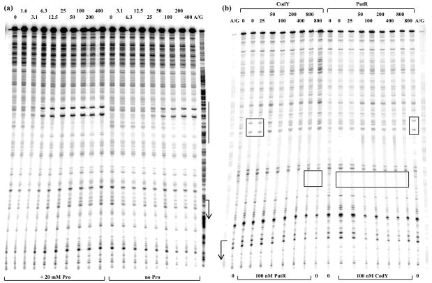
Proline is an efficient source of both carbon and nitrogen for many bacterial species. In Bacillus subtilis, the proline utilization pathway, encoded by the putBCP operon, is inducible by proline. Here, we show that this induction is mediated by PutR, a proline-responsive transcriptional activator of the PucR family. When other amino acids are present in the medium, proline utilization is prioritized through transient repression by CodY, a global transcriptional regulator in Gram-positive bacteria that responds to amino acid availability. CodY-mediated repression of the putBCP operon has two novel features. First, repression requires the cooperative binding of CodY to at least two adjacent motifs. Second, though CodY binds to the region that overlaps the putB promoter, repression is due to displacement of PutR rather than competition with RNA polymerase.


Figures

Fig. 1
Fig. 1
Schematic map of the putBCPR chromosomal locus and the putB regulatory region. The locations of the transcription start point and predicted transcription terminator of the putBCP operon are indicated by the bent arrow and inverted dotted triangle, respectively. The putative promoters of the putP and putR genes are indicated by dotted bent arrows. The PutR-binding site and CodY-binding motifs are shown as rectangles above and below the line, respectively. The coordinates indicate the boundaries of the putB210-lacZ fusion with respect to the transcription start point. The vertical arrow indicates the position where the putB210-lacZ fusion was truncated to create the putB100-lacZ fusion.
Fig. 2
Fig. 2. Binding of CodY to the putB regulatory region
Radioactively labeled putB210p+ (a), putB210p2 (b), putB210p4 (c), putB100p+ (d), and putR (e) DNA fragments were incubated with increasing amounts of purified CodY in the presence of 10 mM ILV and 2 mM GTP. CodY monomer concentrations used (nM) are indicated below each lane. The arrows indicate complexes in which CodY is likely bound to only one motif, as opposed to two motifs simultaneously. KD, the apparent equilibrium dissociation constant, reflects the protein concentration needed to shift 50% of DNA fragments under conditions of vast protein excess over DNA.
Fig. 3
Fig. 3. Binding of PutR to the putB regulatory region
Radioactively labeled putB210p+ (a, b), putB210p6 (c), putB100p+ (d), and putR (e) DNA fragments were incubated with increasing concentrations of purified PutR. All reactions and the gel and gel-running buffer, other than for part (b), contained 20 mM Pro (no ILV or GTP was included in any of the experiments). PutR monomer concentrations used (nM) are indicated below each lane. KD was defined as in Fig. 3.
Fig. 4
Fig. 4. In vitro transcription from the putB promoter
The putB210p+ PCR fragment was transcribed at 37°C for 20 min using purified B. subtilis σA-containing RNA polymerase holoenzyme. (a) All reactions contained 20 mM Pro and increasing amounts of PutR. PutR monomer concentrations used (nM) are indicated below each lane. (b) Reactions contained no PutR or 100 nM PutR and various concentrations of Pro as indicated below each lane. (c) The putB210p+ PCR fragment was preincubated at 37°C first with RNA polymerase and with or without 100 nM PutR or CodY for 15 min (as indicated above the lanes) and then incubated for additional 15 min in the presence of increasing amounts of either CodY or PutR (nM of monomer as indicated below each lane). Finally, transcription was initiated by addition of the nucleotide mixture. All reactions contained 20 mM Pro, 10 mM ILV, and 0.2 mM GTP.
Fig. 5
Fig. 5. Determination of the putB transcription start point and CodY-binding region
(a) Primer extension analysis of putB mRNA. Primer oBB102 annealing to the lacZ gene of the putB-lacZ fusion was extended with reverse transcriptase using as template total RNA from fusion-containing strains BB3328 (wt) and BB3336 (codY) grown in the 16-amino acid-containing medium. The A + G sequencing ladders obtained for the temp late strand of a putB PCR-generated fragment primed with labeled oBB102 are shown to the right and to the left. The apparent transcription start site of the putB gene is in boldface and marked by the +1 notation. The bent arrow indicates the direction of transcription. (b) DNase I footprinting analysis of CodY binding to the putB regulatory region. The putB210p+ DNA fragment labeled on the tem plate strand was incubated with increasing amounts of purified CodY in the presence of 10 mM ILV and 2 mM GTP and then with DNase I. The corresponding A + G sequencing ladder is shown to the right. The apparent transcription start site and direction of putB transcription are shown by the bent arrow. The protected areas are indicated by the vertical lines. CodY monomer concentrations used (nM) are indicated above each lane. (c) The sequence of the coding (non-template) strand of the putB regulatory region used to construct the putB210-lacZ fusions. The likely initiation codon, −10 and −35 promoter regions, the transcription start site and the position of the putBp6 mutation are in bold face. The direction of transcription and translation is indicated by the arrows. The CodY-binding motifs are boxed. The sequences protected by CodY on the template strand in DNase I footprinting experiments are underlined. The sequences protected by PutR are italicized. The coordinates of the 5′ and 3′ ends of the sequence with respect to the transcription start point are shown in parentheses. The boundaries of DNA fragments used to construct various lacZ fusions are indicated by vertical arrows.
Fig. 6
Fig. 6. Mapping of the PutR-binding region and analysis of competition between CodY and PutR
(a) DNase I footprinting analysis of PutR binding to the putB regulatory region. The putB210p+ DNA fragment labeled on the template strand was incubated with increasing amounts of purified PutR in the presence or absence of 20 mM Pro and then with DNase I. The corresponding A + G sequencing ladder is shown to the right. The apparent transcription start site and direction of putB transcription are shown by the bent arrow. The protected area is indicated by the vertical line. PutR monomer concentrations used (nM) are indicated above each lane. (b) CodY dispaces PutR from the putB regulatory region. The putB210p+ DNA fragment labeled on the template strand was preincubated for 15 min with or without 100 nM PutR or 100 nM CodY in the presence of 20 mM Pro, 10 mM ILV, and 2 mM GTP, then incubated for additional 15 min with increasing amounts of either CodY or PutR, and finally treated with DNase I. The corresponding A + G sequencing ladder is shown to the left and to the right. The apparent transcription start site and direction of putB transcription are shown by the bent arrow. Parts of the protected areas characteristic for PutR or CodY binding are indicated by rectangles. For PutR, the boxed region corresponds to two hypersensitive sites within a protected region. Protein concentrations (nM of monomer) used in the first and the second stages of the incubation are indicated below or above each lane, respectively.
Fig. 7
Fig. 7
Induction of the CodY-repressed putB promoter. Cells containing the putB210p+-lacZ fusion (strain BB3328) were pregrown in the medium containing the 16-amino acid mixture but without Pro and arginine. The culture was split at the moment indicated by an arrow, and Pro was added to half of the culture. β-Galactosidase activity was expressed in Miller units. Diamonds – no Pro, squares – plus Pro.
Fig. 8
Fig. 8. Alignment of putB regulatory regions from different Bacillus species
Nucleotides sequences upstream of and including the translation initiation codons of the putB homologs from seven Bacillus strains were aligned using the CLUSTAL W2.1 software (http://www.ebi.ac.uk/Tools/msa/clustalw2/). Identical nucleotides are indicated by asterisks. Initiation codons and putative −10 and −35 promoter regions of B. subtilis putB are in bold. The CodY-binding motifs are underlined and the dyad-symmetry sequence within the PutR binding site is shown by arrows. The accession numbers for the sequences analyzed are: B. subtilis subsp. subtilis (B.s.sub.) - AL009126.3, B. subtilis BSn5 (B.s.Bsn5) - CP002468.1, B. subtilis subsp. spizizenii (B.s.sp.) - CP002183.1, B. amyloliquefaciens (B.amyl.) -FN597644.1, B. atrophaeus (B.atr.) - CP002207.1, B. licheniformis (B.lich.) - NC_006270, B. pumilus (B.pum.) - NC_009848.1. The coordinates are with respect to the translation initiation codons.

Similar articles

Cited by

References

    1. Slack FJ, Serror P, Joyce E, Sonenshein AL. A gene required for nutritional repression of the Bacillus subtilis dipeptide permease operon. Mol Microbiol. 1995;15:689–702. - PubMed
    1. Molle V, Nakaura Y, Shivers RP, Yamaguchi H, Losick R, Fujita Y, Sonenshein AL. Additional targets of the Bacillus subtilis global regulator CodY identified by chromatin immunoprecipitation and genome-wide transcript analysis. J Bacteriol. 2003;185:1911–1922. - PMC - PubMed
    1. Sonenshein AL. CodY, a global regulator of stationary phase and virulence in Gram-positive bacteria. Curr Opin Microbiol. 2005;8:203–207. - PubMed
    1. Sonenshein AL. Control of key metabolic intersections in Bacillus subtilis. Nat Rev Microbiol. 2007;5:917–927. - PubMed
    1. Fisher SH. Regulation of nitrogen metabolism in Bacillus subtilis: vive la difference! Mol Microbiol. 1999;32:223–232. - PubMed

Publication types

MeSH terms

LinkOut - more resources