

How does morphology relate to function in sensory arbors?
- PMID: 21840610
- PMCID: PMC3166259
- DOI: 10.1016/j.tins.2011.07.004
How does morphology relate to function in sensory arbors?
Abstract
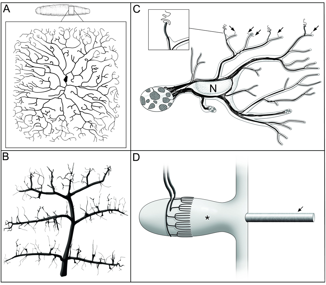
Sensory dendrites fall into many different morphological and functional classes. Polymodal nociceptors are one subclass of sensory neurons, which are of particular note owing to their elaborate dendritic arbors. Complex developmental programs are required to form these arbors and there is striking conservation of morphology, function and molecular determinants between vertebrate and invertebrate polymodal nociceptors. Based on these studies, we argue that arbor morphology plays an important role in the function of polymodal nociceptors. Similar associations between form and function might explain the plethora of dendrite morphologies seen among all sensory neurons.
Copyright © 2011 Elsevier Ltd. All rights reserved.
Figures


Similar articles
-
Neuronal morphogenesis: worms get an EFF in dendritic arborization.Curr Biol. 2010 Aug 24;20(16):R673-5. doi: 10.1016/j.cub.2010.06.053. Curr Biol. 2010. PMID: 20728052
-
The bHLH-PAS protein Spineless is necessary for the diversification of dendrite morphology of Drosophila dendritic arborization neurons.Genes Dev. 2006 Oct 15;20(20):2806-19. doi: 10.1101/gad.1459706. Epub 2006 Oct 2. Genes Dev. 2006. PMID: 17015425 Free PMC article.
-
Dendrite morphogenesis in Caenorhabditis elegans.Genetics. 2024 Jun 5;227(2):iyae056. doi: 10.1093/genetics/iyae056. Genetics. 2024. PMID: 38785371 Free PMC article. Review.
-
Conserved Tao Kinase Activity Regulates Dendritic Arborization, Cytoskeletal Dynamics, and Sensory Function in Drosophila.J Neurosci. 2020 Feb 26;40(9):1819-1833. doi: 10.1523/JNEUROSCI.1846-19.2020. Epub 2020 Jan 21. J Neurosci. 2020. PMID: 31964717 Free PMC article.
-
The Ruffini ending as the primary mechanoreceptor in the periodontal ligament: its morphology, cytochemical features, regeneration, and development.Crit Rev Oral Biol Med. 1999;10(3):307-27. doi: 10.1177/10454411990100030401. Crit Rev Oral Biol Med. 1999. PMID: 10759411 Review.
Cited by
-
Separate transcriptionally regulated pathways specify distinct classes of sister dendrites in a nociceptive neuron.Dev Biol. 2017 Dec 15;432(2):248-257. doi: 10.1016/j.ydbio.2017.10.009. Epub 2017 Oct 13. Dev Biol. 2017. PMID: 29031632 Free PMC article.
-
Caenorhabditis elegans nicotinic acetylcholine receptors are required for nociception.Mol Cell Neurosci. 2014 Mar;59:85-96. doi: 10.1016/j.mcn.2014.02.001. Epub 2014 Feb 8. Mol Cell Neurosci. 2014. PMID: 24518198 Free PMC article.
-
Cellular Pathogenesis of Chemotherapy-Induced Peripheral Neuropathy: Insights From Drosophila and Human-Engineered Skin Models.Front Pain Res (Lausanne). 2022 Jul 8;3:912977. doi: 10.3389/fpain.2022.912977. eCollection 2022. Front Pain Res (Lausanne). 2022. PMID: 35875478 Free PMC article. Review.
-
Precise segmentation of densely interweaving neuron clusters using G-Cut.Nat Commun. 2019 Apr 4;10(1):1549. doi: 10.1038/s41467-019-09515-0. Nat Commun. 2019. PMID: 30948706 Free PMC article.
-
Skin-derived cues control arborization of sensory dendrites in Caenorhabditis elegans.Cell. 2013 Oct 10;155(2):308-20. doi: 10.1016/j.cell.2013.08.058. Cell. 2013. PMID: 24120132 Free PMC article.
References
-
- Ramon y, Cajal S. Histology of the Nervous System of Man and Vertebrate. Oxford University Press: 1995.
-
- Cash S, Yuste R. Linear summation of excitatory inputs by CA1 pyramidal neurons. Neuron. 1999;22:383–394. - PubMed
-
- Branco T, Hausser M. The single dendritic branch as a fundamental functional unit in the nervous system. Curr. Opin. Neurobiol. 2010;20:494–502. - PubMed
Publication types
MeSH terms
Grants and funding
LinkOut - more resources
Full Text Sources
Molecular Biology Databases
