. 2011 Oct;9(10):1418-28.
doi: 10.1158/1541-7786.MCR-11-0147. Epub 2011 Aug 12.

Cross-talk between phospho-STAT3 and PLCγ1 plays a critical role in colorectal tumorigenesis

Affiliations

Cross-talk between phospho-STAT3 and PLCγ1 plays a critical role in colorectal tumorigenesis

Peng Zhang et al. Mol Cancer Res. 2011 Oct.

Abstract

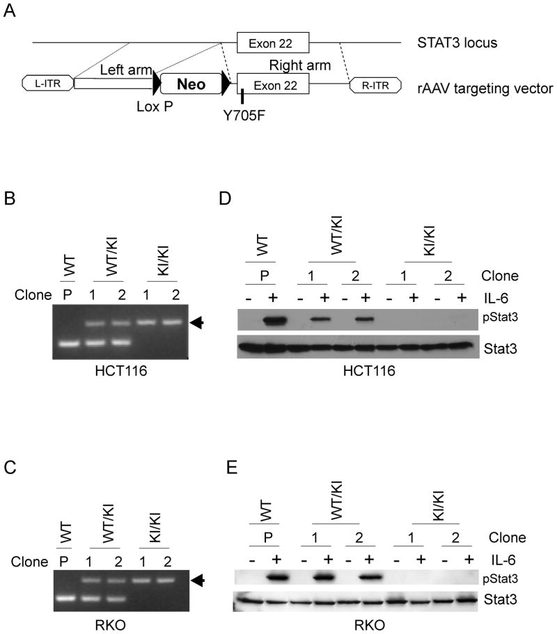
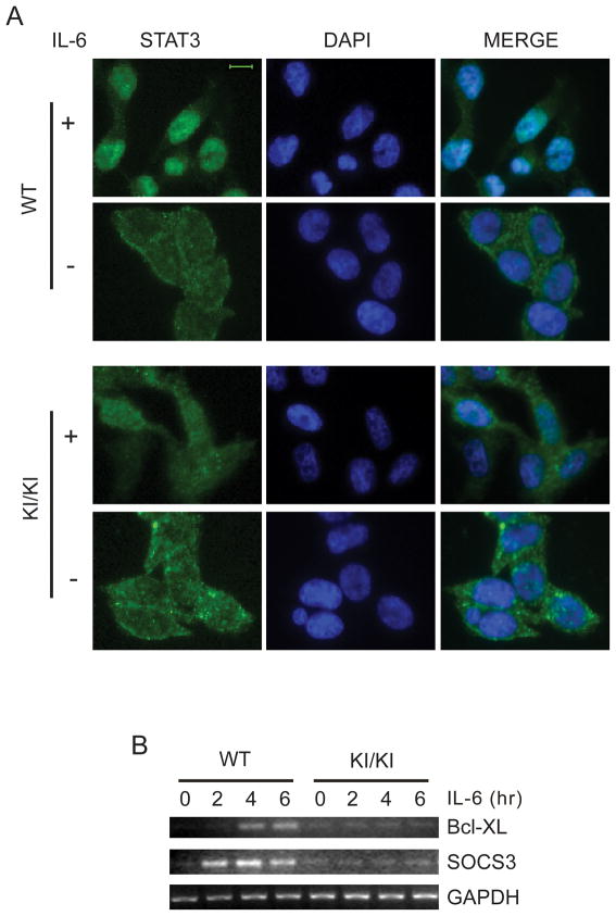
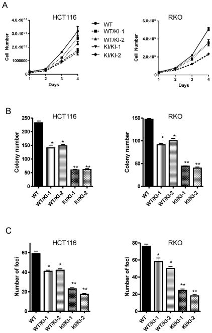
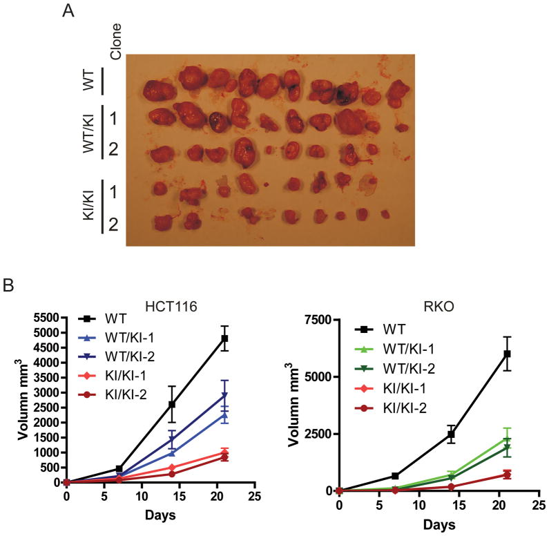
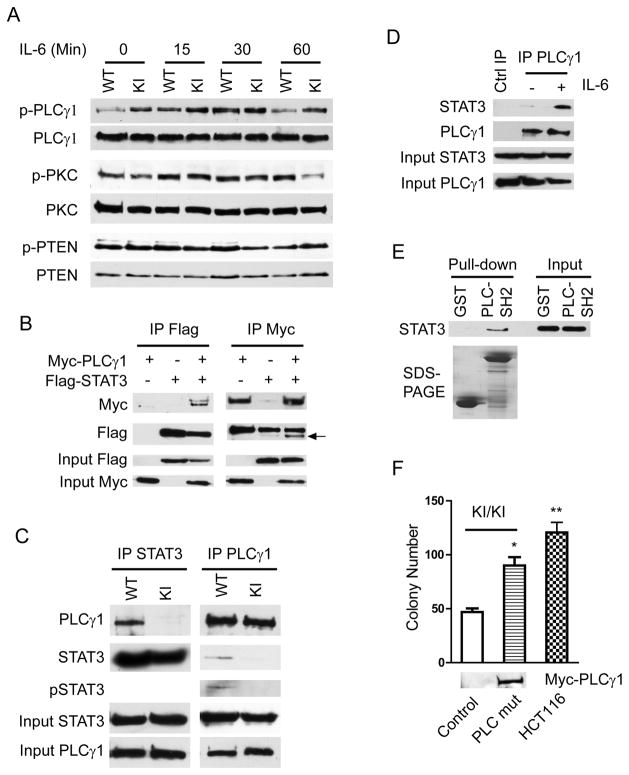
Hyperphosphorylation at the Y705 residue of signal transducer and activator of transcription 3 (STAT3) is implicated in tumorigenesis of leukemia and some solid tumors. However, its role in the development of colorectal cancer is not well defined. To rigorously test the impact of this phosphorylation on colorectal tumorigenesis, we engineered a STAT3 Y705F knock-in to interrupt STAT3 activity in HCT116 and RKO colorectal cancer cells. These STAT3 Y705F mutant cells fail to respond to cytokine stimulation and grow slower than parental cells. These mutant cells are also greatly diminished in their abilities to form colonies in culture, to exhibit anchorage-independent growth in soft agar, and to grow as xenografts in nude mice. These observations strongly support the premise that STAT3 Y705 phosphorylation is crucial in colorectal tumorigenesis. Although it is generally believed that STAT3 functions as a transcription factor, recent studies indicate that transcription-independent functions of STAT3 also play an important role in tumorigenesis. We show here that wild-type STAT3, but not STAT3 Y705F mutant protein, associates with phospholipase Cγ1 (PLCγ1). PLCγ1 is a central signal transducer of growth factor and cytokine signaling pathways that are involved in tumorigenesis. In STAT3 Y705F mutant colorectal cancer cells, PLCγ1 activity is reduced. Moreover, overexpression of a constitutively active form of PLCγ1 rescues the transformation defect of STAT3 Y705F mutant cells. In aggregate, our study identifies previously unknown cross-talk between STAT3 and the PLCγ signaling pathways that may play a critical role in colorectal tumorigenesis.


Figures

Figure 1
Figure 1. Engineering Y705F STAT3 knock-in (KI) colorectal cancer cell lines
(A) Diagram of the KI construct. (B, C) PCR products of the KI clones after excision of the neomycin resistance gene by Cre recombinase. P indicates parental cells; WT/KI indicates Y705F STAT3 heterozygous KI cells; KI/KI indicates Y705F STAT3 homozygous KI cells; arrow indicates the KI allele. (D, E) Parental, Y705F heterozygous and Y705F homozygous cells were starved for 18 hours and treated with or without IL-6 for 30 minutes. Western blots were performed with pSTAT3 or STAT3 antibodies.
Figure 2
Figure 2. STAT3 Y705F mutant proteins fail to activate its target genes
(A) Wild-type and STAT3 Y705F homozygous KI HCT116 CRC cells were starved for 18 hours and stimulated with IL-6 for 30 minutes. Cells were fixed and stained with anti-STAT3 antibodies. Scale bar = 10 μm. (B) Wild-type and STAT3 Y705F homozygous KI HCT116 CRC cells were starved for 18 hours and stimulated with IL-6 for the indicated times. Gene expression of STAT3 target genes, Bcl-XL and SOCS3, were assayed by RT-PCR. GAPDH served as the control.
Figure 3
Figure 3. STAT3 Y705F mutant CRC cells are less tumorigenic in vitro
(A) 1×10⁵ cells of indicated clones were plated in 6-well plates. Duplicate wells of each clone were counted daily for 4 consecutive days using a hemocytometer. Each well was counted three times. Average cell numbers of each time point from three independent experiments are plotted. (B) Cells from indicated clones were plated in 6-well plates in triplicates. Cells were grown for 14 days and stained with crystal violet. Colony numbers were counted and plotted for each of the clones. (C) CRC cells of the indicated clones were mixed in 0.4% soft agar and plated in 6-well plates in triplicates. Cells were grown for 30 days. Colony foci were counted and plotted for each of the clones. * p < 0.05, **p < 0.001, t test.
Figure 4
Figure 4. STAT3 Y705F mutant CRC cells are less tumorigenic in vivo
Athymic nude mice were injected subcutaneously with cells from the indicated clones. Tumors sizes were measured weekly for 3 weeks. Mice were then sacrificed and tumors were harvested. (A) Tumors grown from HCT116 clones. (B) Average sizes of the indicated clones were plotted.
Figure 5
Figure 5. STAT3 cross-talks with PLCγ1
(A) Parental and STAT3 Y705F KI cells were stimulated with IL-6 for the indicated time. Cell lysates were blotted with the indicated antibodies. See Fig. S5 for quantification. (B) HEK 293 cells were transfected with the indicated plasmids. Cell lysates were immunoprecipitated with either anti-Flag or anti-Myc antibodies and blotted with the indicated antibodies. Arrow indicates co-immunoprecipitated Flag-STAT3. (C) Cell lysates from parental and STAT3 Y705F KI cells were immunoprecipitated with antibodies against either STAT3 or PLCγ1 and blotted with the indicated antibodies. (D). HCT116 cells were serum-starved for 16 hours and then stimulated with or without 10 ng/ml of IL-6 for 30 minutes. Cells were then lysed and immunoprecipitated with either anti-PLCγ1 antibodies or control IgG. Immunocomplexes were resolved on 8% SDS-PAGE and Western blots were performed with the indicated antibodies. (E) HCT116 cell lysates were incubated with 1 μg of either GST-PLC γ1-SH2 domains or GST alone for 1 hour at 4°C. The pull-down mixtures were resolved on SDS-PAGE and blotted with an anti-STAT3 antibody. (F) STAT3 Y705F KI cells were transfected with either an empty vector or a vector expressing PLC γ1 triple mutant (D1019L, Y509A and F510A). Colony formation assay was performed. *p < 0.05, ** p < 0.001, t test. Western blot was performed to show expression of myc-tagged PLC γ1 mutant protein.

Similar articles

Cited by

References

    1. Levy DE, Inghirami G. STAT3: a multifaceted oncogene. Proc Natl Acad Sci U S A. 2006;103:10151–2. - PMC - PubMed
    1. Bowman T, Garcia R, Turkson J, Jove R. STATs in oncogenesis. Oncogene. 2000;19:2474–88. - PubMed
    1. Bromberg JF, Wrzeszczynska MH, Devgan G, Zhao Y, Pestell RG, Albanese C, et al. Stat3 as an oncogene. Cell. 1999;98:295–303. - PubMed
    1. Bromberg JF, Horvath CM, Besser D, Lathem WW, Darnell JE., Jr Stat3 activation is required for cellular transformation by v-src. Mol Cell Biol. 1998;18:2553–8. - PMC - PubMed
    1. Wegrzyn J, Potla R, Chwae YJ, Sepuri NB, Zhang Q, Koeck T, et al. Function of mitochondrial Stat3 in cellular respiration. Science. 2009;323:793–7. - PMC - PubMed

Publication types