. 2011 Oct;193(20):5802-9.
doi: 10.1128/JB.05299-11. Epub 2011 Aug 12.

Lipoarabinomannan localization and abundance during growth of Mycobacterium smegmatis

Affiliations

Lipoarabinomannan localization and abundance during growth of Mycobacterium smegmatis

Rakesh K Dhiman et al. J Bacteriol. 2011 Oct.

Abstract

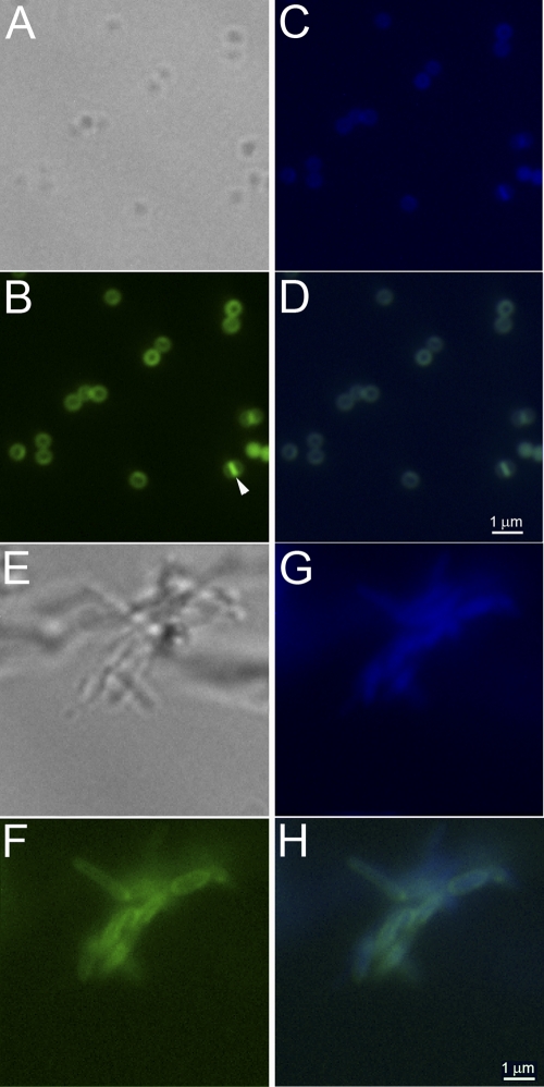
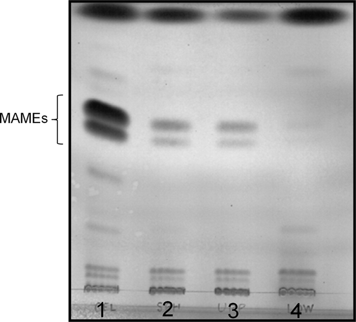
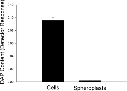
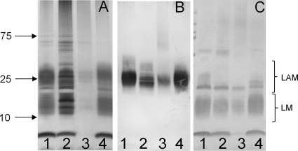
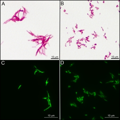
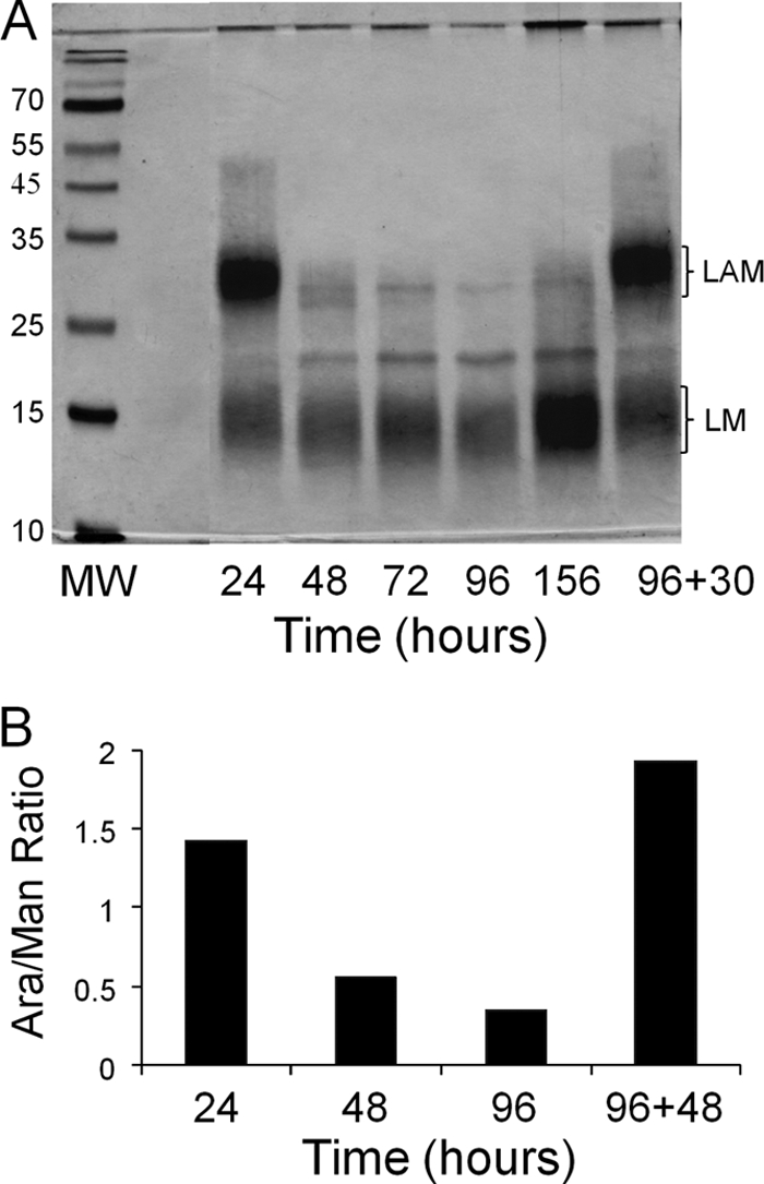
Lipoarabinomannan (LAM) is a structurally heterogeneous amphipathic lipoglycan present in Mycobacterium spp. and other actinomycetes, which constitutes a major component of the cell wall and exhibits a wide spectrum of immunomodulatory effects. Analysis of Mycobacterium smegmatis subcellular fractions and spheroplasts showed that LAM and lipomannan (LM) were primarily found in a cell wall-enriched subcellular fraction and correlated with the presence (or absence) of the mycolic acids in spheroplast preparations, suggesting that LAM and LM are primarily associated with the putative outer membrane of mycobacteria. During the course of these studies significant changes in the LAM/LM content of the cell wall were noted relative to the age of the culture. The LAM content of the M. smegmatis cell wall was dramatically reduced as the bacilli approached stationary phase, whereas LM, mycolic acid, and arabinogalactan content appeared to be unchanged. In addition, cell morphology and acid-fast staining characteristics showed variations with growth phase of the bacteria. In the logarithmic phase, the bacteria were found to be classic rod-shaped acid-fast bacilli, while in the stationary phase M. smegmatis lost the characteristic rod shape and developed a punctate acid-fast staining pattern with carbolfuchsin. The number of viable bacteria was independent of LAM content and phenotype. Taken together, the results presented here suggest that LAM is primarily localized with the mycolic acids in the cell wall and that the cellular concentration of LAM in M. smegmatis is selectively modulated with the growth phase.


Figures

Fig. 1.
Fig. 1.
Morphology of spheroplasts as visualized by labeling with DAPI- and BODIPY-labeled vancomycin. M. smegmatis mc2155 cells were treated with glycine and lysozyme as described in Materials and Methods to generate spheroplasts. Cellular morphology was observed by light and fluorescence microscopy after labeling with BODIPY-labeled vancomycin and DAPI. Spheroplasts are shown in panels A to D, and wild-type M. smegmatis cells are shown in panels E to H. (A and E) Bright-field images; (B and F) BODIPY-vancomycin-labeled peptidoglycan and precursor images (green); (C and G) DAPI-labeled nucleic acid images (blue); (D and H) BODIPY-vancomycin-labeled peptidoglycan and DAPI-labeled nucleic acid images overlaid. The arrowhead in panel B indicates a spheroplast with a visible septum.
Fig. 2.
Fig. 2.
MAME analysis of spheroplast preparations. MAMEs were extracted from equal amounts (by dry weight) of untreated cells or spheroplast preparations and analyzed on silica gel 60 TLC plates developed four times with hexane-ethyl acetate (95:5). MAMEs were visualized by charring. Lane 1, untreated M. smegmatis cells; lane 2, total spheroplasts; lane 3, low-density spheroplast preparation; lane 4, high-density spheroplast preparation.
Fig. 3.
Fig. 3.
Comparison of the DAP content of M. smegmatis cells and spheroplasts. The analysis was normalized to dry weight; the y axis scale is detector response (area units of the peak identified as DAP) per mg (dry weight) of cells or spheroplasts. The values shown are averages of triplicate measurements, and the error bars indicate the standard deviations.
Fig. 4.
Fig. 4.
LAM and LM analysis of control cells and spheroplast preparation. LAM and LM were extracted from equivalent amounts (by dry weight) of untreated M. smegmatis cells and spheroplast preparation. The samples were subjected to SDS-PAGE gel analysis visualized by silver staining (A) or by probing electroblots with CS-35 (B) or conjugated ConA (C). Lane 1, untreated cells in mid-log phase; lane 2, cells treated with glycine only; lane 3, cells treated with glycine and lysozyme (spheroplasts); lane 4, spheroplasts subcultured and regrown for 6 days.
Fig. 5.
Fig. 5.
Growth-phase-associated changes in the morphology and acid-fast staining of M. smegmatis. M. smegmatis mc2155 cells were grown in 7H9 medium supplemented with OADC and 0.05% Tween 80 at 37°C. (A and B) Cells grown for 24 h (A) or 144 h (B) and subjected to Ziehl-Neelsen acid-fast staining without methylene blue counterstaining and light microscopy. (C and D) Cells grown for 24 h (C) or 144 h (D) and subjected to a proprietary acid-fast nucleic acid stain and fluorescence microscopy.
Fig. 6.
Fig. 6.
LM and LAM analysis (A) and the Ara/Man ratio of M smegmatis mc2155 (B). M. smegmatis cells were grown in 7H9 medium supplemented with OADC and 0.05% Tween 80. (A) SDS-PAGE analysis of LAM and LM extracted from cells removed from culture at the indicated time points. The material in the lane labeled 96+30 was from an independent aliquot of cells from the 96-h culture that was diluted 1:100 in fresh medium, followed by incubation at 37°C for an additional 30 h. In all cases, the cells were harvested by centrifugation, dried, weighed, and extracted. Extract from equivalent amounts of cell mass was loaded in each lane. (B) Ratio of arabinose to mannose in the LAM and LM extract from cells removed from culture at the indicated time points. The value indicated by the bar labeled 96+48 was from an independent aliquot of cells from the 96-h culture that was diluted 1:100 in fresh medium, followed by incubation at 37°C for an additional 48 h. The analysis, carried out as described in Materials and Methods, is representative of at least three independent experiments.

Similar articles

Cited by

References

    1. Alderwick L. J., Birch H. L., Mishra A., Eggeling L., Besra G. S. 2007. Structure, function and biosynthesis of the Mycobacterium tuberculosis cell wall: arabinogalactan and lipoarabinomannan assembly with a view to discovering new drug targets. Biochem. Soc. Trans. 35:1325–1328 - PubMed
    1. Alsteens D., et al. 2008. Organization of the mycobacterial cell wall: a nanoscale view. Eur. J. Physiol. 456:117–125 - PubMed
    1. Astarie-Dequeker C., et al. 1999. The mannose receptor mediates uptake of pathogenic and nonpathogenic mycobacteria and bypasses bactericidal responses in human macrophages. Infect. Immun. 67:469–477 - PMC - PubMed
    1. Besra G. S. 1998. Preparation of cell wall fractions from mycobacteria. Methods Mol. Biol. 101:91–107 - PubMed
    1. Betts J. C., Lukey P. T., Robb L. C., McAdam R. A., Duncan K. 2002. Evaluation of a nutrient starvation model of Mycobacterium tuberculosis persistence by gene and protein expression profiling. Mol. Microbiol. 43:717–731 - PubMed

Publication types

MeSH terms

LinkOut - more resources