. 2011 Oct;193(20):5692-706.
doi: 10.1128/JB.05424-11. Epub 2011 Aug 12.

The biofilm inhibitor carolacton disturbs membrane integrity and cell division of Streptococcus mutans through the serine/threonine protein kinase PknB

Affiliations

The biofilm inhibitor carolacton disturbs membrane integrity and cell division of Streptococcus mutans through the serine/threonine protein kinase PknB

Michael Reck et al. J Bacteriol. 2011 Oct.

Abstract

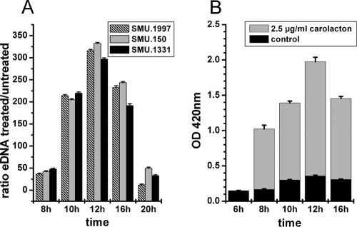
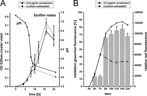
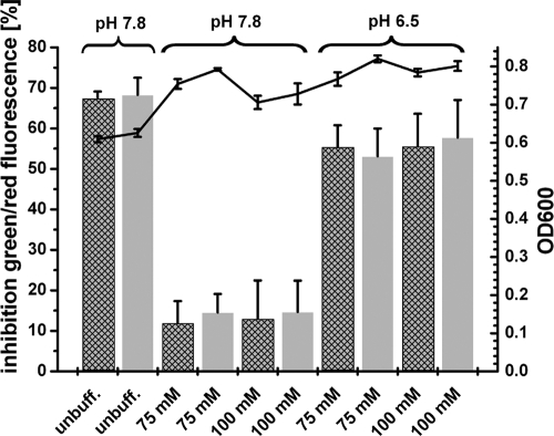
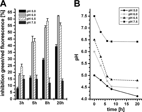
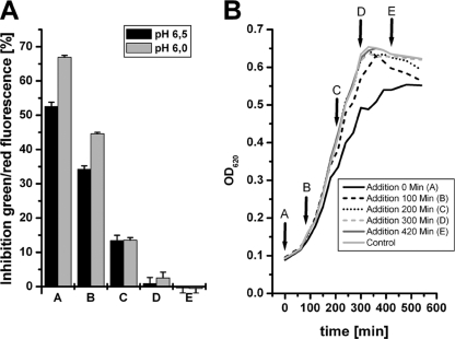
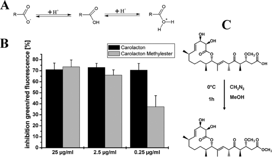
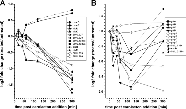
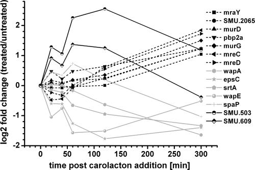
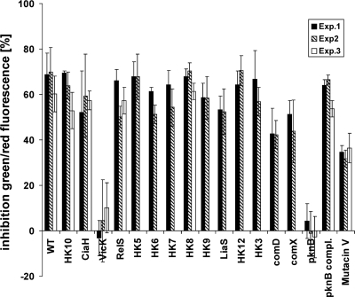
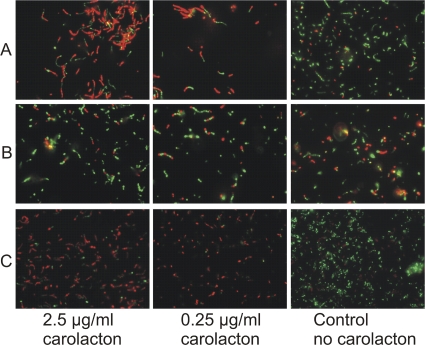
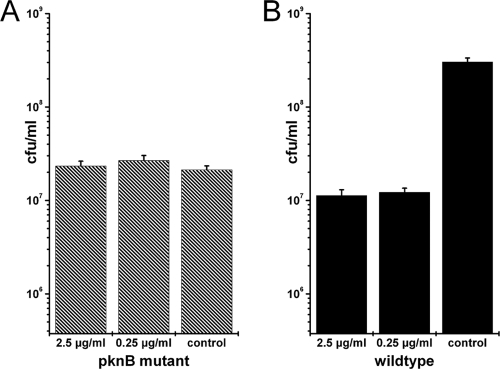
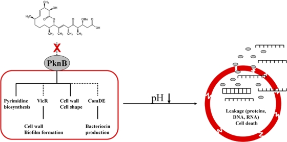
Carolacton, a secondary metabolite isolated from the myxobacterium Sorangium cellulosum, disturbs Streptococcus mutans biofilm viability at nanomolar concentrations. Here we show that carolacton causes leakage of cytoplasmic content (DNA and proteins) in growing cells at low pH and provide quantitative data on the membrane damage. Furthermore, we demonstrate that the biofilm-specific activity of carolacton is due to the strong acidification occurring during biofilm growth. The chemical conversion of the ketocarbonic function of the molecule to a carolacton methylester did not impact its activity, indicating that carolacton is not functionally activated at low pH by a change of its net charge. A comparative time series microarray analysis identified the VicKRX and ComDE two-component signal transduction systems and genes involved in cell wall metabolism as playing essential roles in the response to carolacton treatment. A sensitivity testing of mutants with deletions of all 13 viable histidine kinases and the serine/threonine protein kinase PknB of S. mutans identified only the ΔpknB deletion mutant as being insensitive to carolacton treatment. A strong overlap between the regulon of PknB in S. mutans and the genes affected by carolacton treatment was found. The data suggest that carolacton acts by interfering with PknB-mediated signaling in growing cells. The resulting altered cell wall morphology causes membrane damage and cell death at low pH.


Figures

Fig. 1.
Fig. 1.
Carolacton causes leakage of cytoplasmic DNA (A) and proteins (B). (A) Relative quantification of the external DNA (eDNA) of carolacton-treated biofilms. Supernatants of carolacton-treated (2.5 μg/ml) and untreated biofilm cells were analyzed using quantitative PCRs specific for 3 different genes randomly distributed over the S. mutans genome. The means and standard deviations from 3 biological replicates are shown. (B) Carolacton-caused release of intracellular β-galactosidase into the supernatant during biofilm growth of an S. mutans reporter strain (MR15 [Table 1]). The β-galactosidase activity was measured by hydrolysis of the β-galactosidase substrate ortho-nitrophenyl-β-d-galactopyranoside (ONPG) to galactose and o-nitrophenol in the supernatant of carolacton-treated and untreated biofilm cells. The OD420, corresponding to the absorption maximum of o-nitrophenol, is plotted against the time of biofilm growth. Means and standard deviations from 3 biological replicates are shown.
Fig. 2.
Fig. 2.
Effect of carolacton on biofilm mass (A) and viability (B) and the corresponding pH drop. (A) Biofilm mass and pH during growth of carolacton-treated and untreated samples, as determined by crystal violet staining. The means and the standard deviations from two biological replicates, each consisting of 6 technical replicates, are presented. (B) Inhibition of green/red fluorescence (bar chart), as a measure for viability, and the red fluorescence (line plots), as a measure for membrane damage, for LIVE/DEAD-stained biofilms. For the inhibition of green/red fluorescence, the means and standard deviations from 2 biological replicates, each consisting of 6 technical replicates, are shown.
Fig. 3.
Fig. 3.
Carolacton activity depends on pH. Carolacton was added to phosphate-buffered (75 and 100 mM) and unbuffered (unbuff.) THBS media before inoculation with S. mutans UA159 to an OD600 of 0.01. Two different carolacton concentrations were tested, 0.25 μg/ml (crosshatched bars) and 2.5 μg/ml (gray bars). The media differed in buffer concentration (75 mM versus 100 mM) and in the initial pH (7.8 versus 6.5). After 20 h, biofilms were washed and stained with LIVE/DEAD viability staining. The inhibition of viability was calculated relative to untreated control wells (see Materials and Methods). Control wells contain the same amount of methanol as used for the carolacton-treated wells but lacked carolacton. Standard deviations were calculated from 3 independent biological replicates, each consisting of 5 technical replicates. The mean of the OD600 is shown as a line graph.
Fig. 4.
Fig. 4.
Sensitivity to carolacton of planktonic cells growing under acidic conditions. Carolacton-treated (2.5 μg/ml) planktonic cells growing in buffered (75 mM phosphate buffer) THBY medium at different initial pH values were cultivated under anaerobic conditions. (A) Inhibition of viability as determined by LIVE/DEAD staining. (B) pH in the cultures during growth.
Fig. 5.
Fig. 5.
Role of growth phase in the membrane damage caused by carolacton in planktonic culture. Carolacton (2.5 μg/ml) was added at different growth phases (conditions A to E) to planktonic cultures of S. mutans growing in buffered (75 mM phosphate buffer) THBY medium at pH 6.0. and 6.5. (A) Inhibition of viability determined by LIVE/DEAD viability staining. (B) Growth of planktonic cells at pH 6.0.
Fig. 6.
Fig. 6.
Influence of the carboxylic moiety on carolacton activity. (A) Change of the charge of the ketocarbonic function of carolacton upon decreasing pH. (B) Inhibition of biofilm viability by carolacton and the carolacton methylester for 3 different inhibitor concentrations as determined by LIVE/DEAD viability staining. (C) Conversion of carolacton into the corresponding methylester.
Fig. 7.
Fig. 7.
Effect of carolacton on the expression of TCSs of S. mutans (A) and VicKRX-regulated genes (B) as determined by microarray time series analysis. (A) The differential expression profiles of the response regulators are marked with solid lines, while those of the cognate histidine kinases are marked with dotted lines. (B) Relative expression of genes known to be regulated by the VicKRX system during 300 min of carolacton treatment.
Fig. 8.
Fig. 8.
Carolacton influences expression of genes involved in cell wall metabolism and cell division. The relative expression profiles for genes coding for membrane proteins (SMU.503 and SMU.609), proteins involved in cell wall metabolism and cell division (MreCD, SMU.2065, MraY, MurDG, and Pbp2a), and surface proteins (SrtA, WapAE, and SpaP), determined by microarray analysis, are shown.
Fig. 9.
Fig. 9.
Sensitivity to carolacton of mutants with deletions of potential target genes. Mutants of S. mutans deficient for the 13 viable HKs (HK3, HK5 to -10, HK12, LiaS, VicK, CiaH, RelS, and ComD), mutacin V synthesis (Mutacin V), the serine/threonine membrane kinase PknB, and the alternative sigma factor ComX were tested for their sensitivity to carolacton using LIVE/DEAD viability staining of biofilms. The means and standard deviations from at least 5 technical replicates are shown. Each experiment was performed in 2 or 3 biological replicates.
Fig. 10.
Fig. 10.
Verification of the insensitivity of a pknB-deficient strain to carolacton using LIVE/DEAD staining and fluorescence microscopy. Images of carolacton-treated and untreated biofilm cells of the wild type (A), the ΔpknB mutant (B), and the complemented strain (C) are shown.
Fig. 11.
Fig. 11.
Verification of the insensitivity of a pknB mutant to carolacton by determination of CFU. CFU of carolacton-treated (2.5 μg/ml or 0.25 μg/ml) and untreated (control) biofilm cells of a pknB-deficient strain (A) and the wild type (B) are shown. Cells derived from 3 different wells of the microtiter plate were diluted independently, and each dilution was plated out on 3 agar plates, resulting in 9 different values for each condition. The standard deviation from these 9 technical replicates is shown. Each experiment was repeated twice, and the figure shows a representative result.
Fig. 12.
Fig. 12.
Working model for the mode of action of carolacton. Carolacton is postulated to interfere with PknB-mediated signaling. Dashed lines represent hypothetical interactions. Disturbance of cell wall metabolism by altered PknB signaling might be mediated either directly by altered phosphorylation of target proteins or indirectly via the VicR cascade. Induction of ComDE is accompanied by an upregulation of bacteriocins and bacteriocin-associated genes. Furthermore, repression of pyrimidine biosynthesis occurs, as STPKs are global regulators of gene expression. Alterations in cell wall composition result in a weakened cell wall, leading to loss of membrane integrity at low pH and leakage of the cytoplasmic contents.

Similar articles

Cited by

References

    1. Ahn S. J., Burne R. A. 2006. The atlA operon of Streptococcus mutans: role in autolysin maturation and cell surface biogenesis. J. Bacteriol. 188:6877–6888 - PMC - PubMed
    1. Ahn S. J., Burne R. A. 2007. Effects of oxygen on biofilm formation and the AtlA autolysin of Streptococcus mutans. J. Bacteriol. 189:6293–6302 - PMC - PubMed
    1. Banu L. D., et al. 2010. The Streptococcus mutans serine/threonine kinase, PknB, regulates competence development, bacteriocin production, and cell wall metabolism. Infect. Immun. 78:2209–2220 - PMC - PubMed
    1. Beltramini A. M., Mukhopadhyay C. D., Pancholi V. 2009. Modulation of cell wall structure and antimicrobial susceptibility by a Staphylococcus aureus eukaryote-like serine/threonine kinase and phosphatase. Infect. Immun. 77:1406–1416 - PMC - PubMed
    1. Biswas I., Jha J. K., Fromm N. 2008. Shuttle expression plasmids for genetic studies in Streptococcus mutans. Microbiology 154:2275–2282 - PMC - PubMed

Publication types

MeSH terms