. 2011 Oct 7;286(40):34809-19.
doi: 10.1074/jbc.M111.281626. Epub 2011 Aug 12.

Repressive chromatin affects factor binding at yeast HO (homothallic switching) promoter

Affiliations

Repressive chromatin affects factor binding at yeast HO (homothallic switching) promoter

Shinya Takahata et al. J Biol Chem. .

Abstract

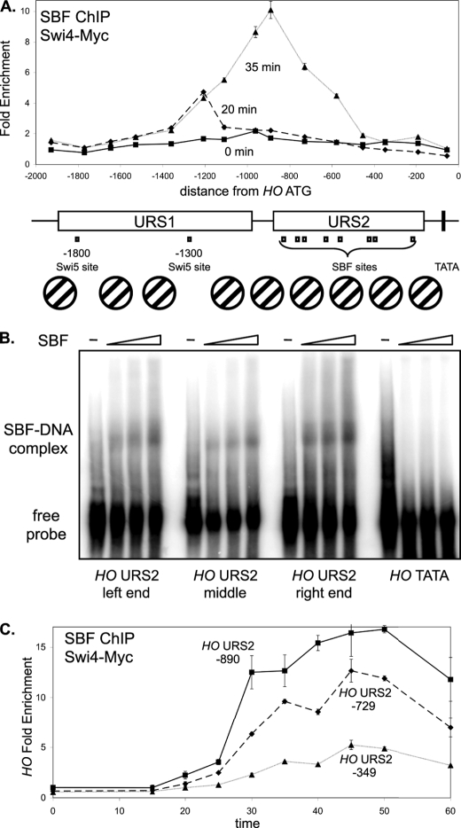
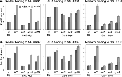
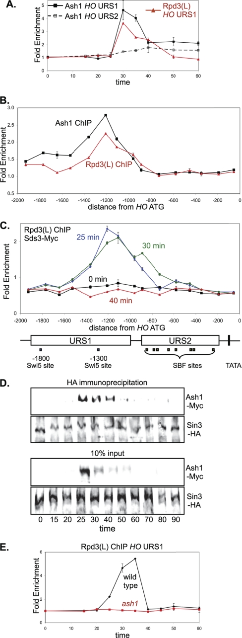
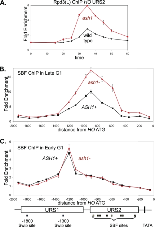
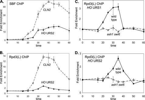
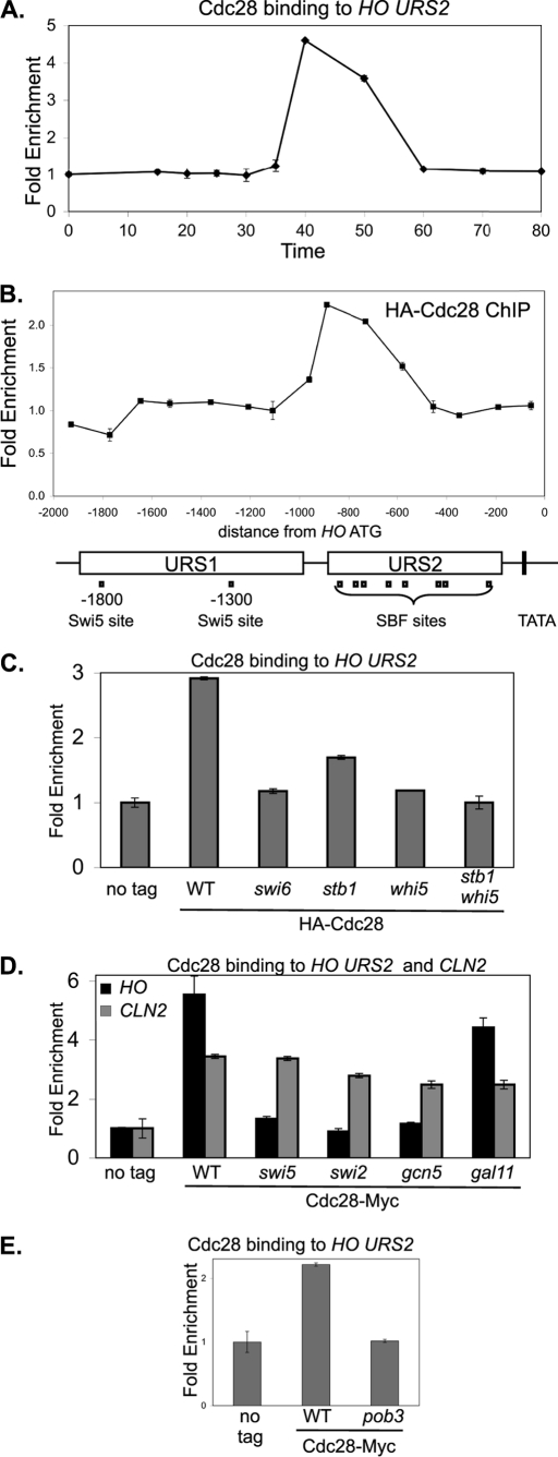
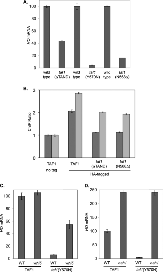
The yeast HO gene is tightly regulated, with multiple activators and coactivators needed to overcome repressive chromatin structures that form over this promoter. Coactivator binding is strongly interdependent, as loss of one factor sharply reduces recruitment of other factors. The Rpd3(L) histone deacetylase is recruited to HO at two distinct times during the cell cycle, first by Ash1 to the URS1 region of the promoter and then by SBF/Whi5/Stb1 to URS2. SBF itself is localized to only a subset of its potential binding sites in URS2, and this localization takes longer and is less robust than at other SBF target genes, suggesting that binding to the HO promoter is limited by chromatin structures that dynamically change as the cell cycle progresses. Ash1 only binds at the URS1 region of the promoter, but an ash1 mutation results in markedly increased binding of SBF and Rpd3(L) at URS2, some 450 bp distant from the site of Ash1 binding, suggesting these two regions of the promoter interact. An ash1 mutation also results in increased coactivator recruitment, Swi/Snf and Mediator localization in the absence of the normally required Gcn5 histone acetyltransferase, and HO expression even in the presence of a taf1 mutation affecting TFIID activity that otherwise blocks HO transcription. Ash1 therefore appears to play a central role in generating the strongly repressive environment at the HO promoter, which limits the binding of several coactivators at URS2 and TATA region.


Figures

FIGURE 1.
FIGURE 1.
SBF binding is strongest to sites at the left end of URS2. A, DY12794 cells (GALp::CDC20 SWI4-Myc) with a GALp::CDC20 allele were synchronized in mitosis by removing galactose, followed by release by addition of galactose (t = 0). The CDC20 arrest is at the G2/M transition, and HO expression at 40 min following release corresponds to late G1 phase. SBF binding was measured by Swi4-Myc ChIP using samples taken at t = 0, 20, and 35 min following the release. ChIP samples were analyzed with 15 sets of PCR primers across the HO promoter, with an average PCR product size of 208 bp. URS1-, URS2-, Swi5- and SBF-binding sites are shown for the HO promoter, where the ATG represents +1 and the transcription start site is at −20. Nucleosome positions along the HO promoter, determined by micrococcal nuclease mapping and by H3 ChIP (3), are shown. B, electrophoretic mobility shift assays examine in vitro binding of recombinant SBF to sites at the left (−857), middle (−461) and right (−246) parts of URS2, and the TATA region as a negative control. C, DY12794 cells (GALp::CDC20 SWI4-Myc) were synchronized and SBF binding by Swi4-Myc ChIP during the time course to three regions of HO URS2 at −890, −729, and −349, where the numbers refer to the center of the amplified region.
FIGURE 2.
FIGURE 2.
Coactivator mutations reduce coactivator binding to HO. ChIP experiments were performed with logarithmically growing untagged control strains (DY150, wild type; DY4394, ash1; DY161, swi5; DY4846, swi5 ash1; DY12761, swi2(E834K); DY9711, swi2(E834K) ash1; DY5925, gcn5; DY7387, gcn5 ash1; DY5628, gal11; and DY14144, gal11 ash1), SWI2-Myc strains (DY6151, wild type; DY7403, ash1; DY9395, swi5; DY6378, swi5 ash1; DY8738, gcn5; DY8736, gcn5 ash1; DY14147, gal11; and DY14148, gal11 ash1), GCN5-Myc strains (DY7196, wild type; DY7194, ash1; DY6067, swi5; DY14137, swi5 ash1; DY12861, swi2(E834K); DY11278, swi2(E834K) ash1; DY7240, gal11; and DY14140, gal11 ash1), and GAL11-Myc strains (DY6148, wild type; DY6739, ash1; DY6197, swi5; DY6741, swi5 ash1; DY12729, swi2(E834K); DY14186, swi2(E834K) ash1; DY12711, gcn5; DY14142, gcn5 ash1). A, binding to URS1 was measured with primers that amplify from −1429 to −1139. B, binding to URS2 was measured with primers that amplify from −825 to −489.
FIGURE 3.
FIGURE 3.
Ash1 recruits Rpd3(L) to HO URS2. A, strains DY8312 (GALp::CDC20 ASH1-Myc) and DY12247 (GALp::CDC20 SDS3-Myc) were synchronized, and ChIP experiments were performed to measure Ash1 and Rpd3(L) binding to the HO promoter. Binding to URS1 and URS2 was measured with primers that amplify from −1429 to −1139 and from −825 to −489, respectively. Sds3 is a subunit specific to Rpd3(L). B, ChIP samples were prepared from logarithmically growing strains DY8312 (ASH1-Myc) and DY12247 (SDS3-Myc), and binding was analyzed with 15 sets of PCR primers across the HO promoter. C, strain DY12247 (GALp::CDC20 SDS3-Myc) was synchronized, and ChIP samples were taken at various times following release from CDC20 arrest. Binding was analyzed with 15 sets of PCR primers across the HO promoter. URS1-, URS2-, Swi5-, and SBF-binding sites are shown for the HO promoter, where the ATG represents +1 and the transcription start site is at −20. D, strain DY13197 (GALp::CDC20 Ash1-Myc SIN3-HA) was synchronized, and protein samples were taken at various times following release from CDC20 arrest. Sin3-HA was immunoprecipitated with anti-HA antibody and analyzed on Western blots, along with controls corresponding to 10% of the input before immunoprecipitation, and the blots were probed with anti-HA and anti-Myc antibodies. E, strains DY12247 (GALp::CDC20 SDS3-Myc) and DY12251 (GALp::CDC20 SDS3-Myc ash1) were synchronized, and ChIP samples were taken at various times following release from CDC20 arrest. Binding to HO URS1 was measured with primers that amplify from −1429 to −1139.
FIGURE 4.
FIGURE 4.
ash1 mutation results in increased SBF and Rpd3L binding. A, strains DY12247 (GALp::CDC20 SDS3-Myc) and DY12251 (GALp::CDC20 SDS3-Myc ash1) were synchronized, and ChIP samples were taken at various times following release from CDC20 arrest. Rpd3(L) binding to HO URS2 was measured with primers that amplify from −825 to −489. Sds3 is a subunit specific to Rpd3(L). B, strains DY12794 (GALp::CDC20 SWI4-Myc) and DY12795 (GALp::CDC20 SWI4-Myc ash1) were synchronized, and ChIP samples were taken at 35 min following release from CDC20 arrest, when cells are in late G1. SBF binding was analyzed with 15 sets of PCR primers across the HO promoter. C, strains DY12794 (GALp::CDC20 SWI4-Myc) and DY12795 (GALp::CDC20 SWI4-Myc ash1) were synchronized, and ChIP samples were taken at 20 min following release from CDC20 arrest, when cells are in early G1. SBF binding was analyzed with 15 sets of PCR primers across the HO promoter. URS1-, URS2-, Swi5-, and SBF-binding sites are shown for the HO promoter, where the ATG represents +1 and the transcription start site is at −20.
FIGURE 5.
FIGURE 5.
SBF and Rpd3(L) binding to HO URS2 is weak compared with binding at CLN2. A, strain DY12794 (GALp::CDC20 SWI4-Myc) was synchronized, and ChIP samples were taken at various times following release from CDC20 arrest. SBF binding to HO URS2 was measured with primers that amplify from −825 to −489 and binding to CLN2 with primers that amplify from −661 to −379. B, strain DY12247 (GALp::CDC20 SDS3-Myc) was synchronized, and ChIP samples were taken at various times following release from CDC20 arrest. SBF binding to HO URS2 was measured with primers that amplify from −825 to −489 and binding to CLN2 with primers that amplify from −661 to −379. Sds3 is a subunit specific to Rpd3(L). C, strains DY12247 (GALp::CDC20 SDS3-Myc) and DY12830 (GALp::CDC20 SDS3-Myc ash1 swi6) were synchronized, and ChIP samples were taken at various times following release from CDC20 arrest. Rpd3(L) binding to HO URS1 was measured with primers that amplify from −1429 to −1139. D, strains DY12247 (GALp::CDC20 SDS3-Myc) and DY12830 (GALp::CDC20 SDS3-Myc ash1 swi6) were synchronized, and ChIP samples were taken at various times following release from CDC20 arrest. Rpd3(L) binding to HO URS2 was measured with primers that amplify from −825 to −489.
FIGURE 6.
FIGURE 6.
Cdc28 kinase is recruited to HO URS2. A, strain DY6669 (GALp::CDC20 SWI4-Myc) with HA-CDC28 on a YCp-KanMX plasmid was synchronized, and ChIP samples were taken at various times following release from CDC20 arrest. Cdc28 binding to HO URS2 was measured with primers that amplify from −825 to −489. B, ChIP samples prepared from logarithmically growing strain DY150 with either a YCp-KanMX(HA-CDC28) plasmid or an empty YCp-KanMX vector were synchronized, and Cdc28 binding was analyzed with 15 sets of PCR primers across the HO promoter. URS1-, URS2-, Swi5-, and SBF-binding sites are shown for the HO promoter, where the ATG represents +1 and the transcription start site is at −20. C, ChIP experiments were performed with logarithmically growing untagged control strains (DY150, wild type; DY6999, swi6; DY13454, stb1; DY9559, whi5; and DY13640, whi5 stb1) and CDC28-Myc strains (DY13020, wild type; DY13723, swi6; DY13729, stb1; DY13731, whi5; and DY13733, stb1 whi5). Cdc28 binding to HO URS2 was measured with primers that amplify from −825 to −489. D, ChIP experiments were performed with logarithmically growing strains (DY150, wild type; DY161, swi5; DY5270, swi2Δ; DY5925, gcn5; and DY5628, gal11) containing either a YCp-KanMX(HA-CDC28) plasmid or an empty YCp-KanMX vector. Cdc28 binding to HO URS2 was measured with primers that amplify from −825 to −489 and binding to CLN2 with primers that amplify from −661 to −379. E, ChIP experiments were performed with logarithmically growing untagged control strains (DY150, wild type; DY7379, pob3(L78R)) and CDC28-Myc strains (DY13020, wild type; DY13527, pob3(L78R)), grown at 25 °C. Cdc28 binding to HO URS2 was measured with primers that amplify from −825 to −489.
FIGURE 7.
FIGURE 7.
Defect in HO expression caused by a taf1 mutation can be suppressed by whi5 or ash1. A, HO mRNA levels were measured by RT-qPCR from cells growing logarithmically at 33 °C: DY150, wild type; DY10598, taf1TAND); DY11348, taf1Δ YCp(TRP1)-TAF1; DY11349, taf1Δ YCp(TRP1)-TAF1(Y570N); DY12303, taf1Δ YCp(HIS3)-TAF1-HA; and DY12305, taf1Δ YCp(HIS3)-TAF1(N568Δ)-HA. B, ChIP experiments were performed from cells growing logarithmically at 33 °C: DY10204 (taf1Δ YCp(TRP1)-TAF1) and DY10366 (taf1Δ YCp(TRP1)-TAF1 ash1) as untagged controls. YCp(HIS3) plasmids expressing TAF1-HA, TAF1(T657K)-HA, or TAF1(N568Δ)-HA were shuffled into strains DY10204 (ASH1) and DY10366 (ash1), and ChIP experiments were performed with the HA-tagged TAF1 as the only copy in the cell. Taf1 binding to the HO TATA was measured with primers that amplify from −43 to +175. C, HO mRNA levels were measured by RT-qPCR from cells growing logarithmically at 33 °C: DY11348, taf1Δ YCp(TRP1)-TAF1; DY11574, taf1Δ YCp(TRP1)-TAF1 whi5; DY11349, taf1Δ YCp(TRP1)-TAF1(Y570N); and DY11575, taf1Δ YCp(TRP1)-TAF1(Y570N) whi5. D, HO mRNA levels were measured by RT-qPCR from cells growing logarithmically at 33 °C: DY11348, taf1Δ YCp(TRP1)-TAF1; DY11570, taf1Δ YCp(TRP1)-TAF1 ash1; DY11349, taf1Δ YCp(TRP1)-TAF1(Y570N); and DY11571, taf1Δ YCp(TRP1)-TAF1(Y570N) ash1.

Similar articles

Cited by

References

    1. Cairns B. R. (2009) Nature 461, 193–198 - PubMed
    1. Weake V. M., Workman J. L. (2010) Nat. Rev. Genet. 11, 426–437 - PubMed
    1. Takahata S., Yu Y., Stillman D. J. (2009) Mol. Cell 34, 405–415 - PMC - PubMed
    1. Peterson C. L., Herskowitz I. (1992) Cell 68, 573–583 - PubMed
    1. Stillman D. J., Dorland S., Yu Y. (1994) Genetics 136, 781–788 - PMC - PubMed

Publication types

MeSH terms

LinkOut - more resources