. 2011 Oct;301(4):H1519-30.
doi: 10.1152/ajpheart.01080.2010. Epub 2011 Aug 12.

microRNA-210 is upregulated in hypoxic cardiomyocytes through Akt- and p53-dependent pathways and exerts cytoprotective effects

Affiliations

microRNA-210 is upregulated in hypoxic cardiomyocytes through Akt- and p53-dependent pathways and exerts cytoprotective effects

R Kannan Mutharasan et al. Am J Physiol Heart Circ Physiol. 2011 Oct.

Abstract

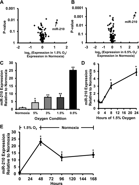
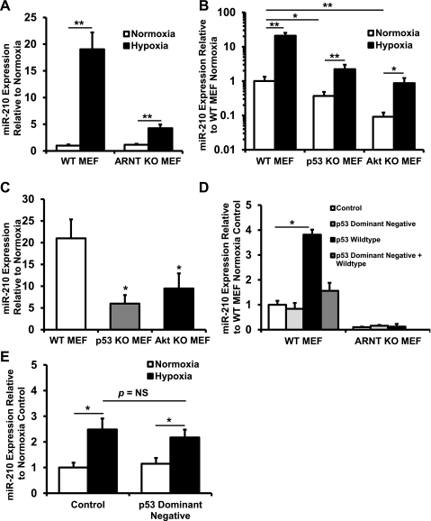
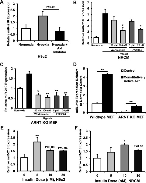
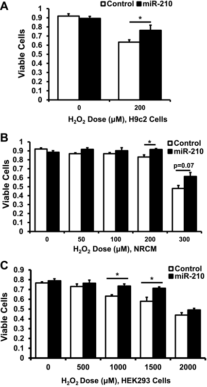
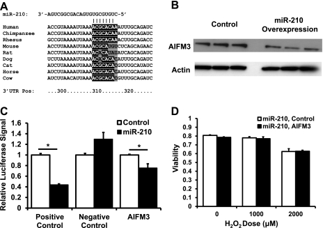
microRNA-210 (miR-210) is upregulated in hypoxia, but its function in cardiomyocytes and its regulation in response to hypoxia are not well characterized. The purpose of this study was to identify upstream regulators of miR-210, as well as to characterize miR-210's function in cardiomyocytes. We first showed miR-210 is upregulated through both hypoxia-inducible factor (HIF)-dependent and -independent pathways, since aryl hydrocarbon nuclear translocator (ARNT) knockout mouse embryonic fibroblasts (MEF), lacking intact HIF signaling, still displayed increased miR-210 levels in hypoxia. To determine the mechanism for HIF-independent regulation of miR-210, we focused on p53 and protein kinase B (Akt). Overexpression of p53 in wild-type MEFs induced miR-210, whereas p53 overexpression in ARNT knockout MEFs did not, suggesting p53 regulates miR-210 in a HIF-dependent mechanism. Akt inhibition reduced miR-210 induction by hypoxia, whereas Akt overexpression increased miR-210 levels in both wild-type and ARNT knockout MEFs, indicating Akt regulation of miR-210 is HIF-independent. We then studied the effects of miR-210 in cardiomyocytes. Overexpression of miR-210 reduced cell death in response to oxidative stress and reduced reactive oxygen species (ROS) production both at baseline and after treatment with antimycin A. Furthermore, downregulation of miR-210 increased ROS after hypoxia-reoxygenation. To determine a mechanism for the cytoprotective effects of miR-210, we focused on the predicted target, apoptosis-inducing factor, mitochondrion-associated 3 (AIFM3), known to induce cell death. Although miR-210 reduced AIFM3 levels, overexpression of AIFM3 in the presence of miR-210 overexpression did not reduce cellular viability either at baseline or after hydrogen peroxide treatment, suggesting AIFM3 does not mediate miR-210's cytoprotective effects. Furthermore, HIF-3α, a negative regulator of HIF signaling, is targeted by miR-210, but miR-210 does not modulate HIF activity. In conclusion, we demonstrate a novel role for p53 and Akt in regulating miR-210 and demonstrate that, in cardiomyocytes, miR-210 exerts cytoprotective effects, potentially by reducing mitochondrial ROS production.


Figures

Fig. 1.
Fig. 1.
microRNA-210 (miR-210) is upregulated in hypoxic cardiomyocytes. A: volcano plot of microRNA expression level and uncorrected P value in the 1.5% O2 for 48 h condition. B: volcano plot of microRNA expression level and uncorrected P value in the 0.5% O2 for 48 h condition. miR-210 is the most strongly induced microRNA in both conditions and, after correction for multiple comparisons, is the only significantly upregulated microRNA (corrected P = 0.04 in the 0.5% O2 condition); n = 3 independent biological samples in each group. C: RT-PCR data confirm that hypoxia upregulates miR-210 in an O2 dose-responsive manner (*P < 0.05 and **P < 0.01). D: miR-210 is induced in neonatal rat cardiomyocytes (NRCM) as soon as 8 h after hypoxia exposure (*P < 0.05 compared with time 0). E: miR-210 levels remain elevated in NRCM up to 120 h after withdrawal of cells from hypoxia (*P < 0.05 compared with time 0). Data are presented as means ± SE; n = 3 in each group.
Fig. 2.
Fig. 2.
miR-210 is regulated through hypoxia-inducible factor (HIF)-dependent and -independent pathways. A: hypoxia (1.5% O2) upregulates miR-210 both in wild-type (WT) and aryl hydrocarbon nuclear translocator (ARNT) knockout (KO) mouse embryonic fibroblasts (MEFs) lacking HIF signaling, a known inducer of miR-210 (**P < 0.01 compared with normoxia, n = 6). The degree of miR-210 induction, however, is lower in ARNT KO MEFs. B: p53 KO MEFs and Akt KO MEFs have lower levels of miR-210 expression both at baseline and in response to hypoxia (*P < 0.05 and **P <0.01 compared with WT MEF; n = 6 in all groups). C: both p53 KO MEFs and Akt KO MEFs have reduced fold increase of miR-210 (*P < 0.05 compared with WT MEF, n = 6 for each group). D: p53 plasmid, but not p53 dominant-negative plasmid, transfected into WT MEFs induces miR-210 expression (*P < 0.05 compared with pCMV control plasmid transfection, n = 3). Transfection of dominant-negative plasmid with WT plasmid abrogates induction of miR-210 by WT p53. However, in ARNT KO MEF, p53 overexpression does not increase miR-210 levels. E: p53 dominant-negative plasmid transfection into ARNT KO MEFs does not reduce induction of miR-210 by hypoxia (*P < 0.05 compared with normoxia, n = 3). NS, not significant. Data are presented as means ± SE.
Fig. 3.
Fig. 3.
Akt upregulates miR-210. A: In H9c2 cells, there was a reduction in miR-210 response to hypoxia after treatment with Akt inhibitor (P = 0.06 compared with hypoxia alone). B: in NRCM, treatment with the phosphatidylinositol 3-kinase (PI 3-kinase) inhibitors wortmannin at 300 nM and LY-29004 at 20 μM prevented induction of miR-210 by hypoxia (*P < 0.05 compared with hypoxia alone). C: in ARNT KO MEFs, treatment with wortmannin and LY-29004 prevented induction of miR-210 by hypoxia (*P < 0.05 and **P < 0.01 compared with hypoxia alone). D: constitutively active Akt plasmid transfection in both WT MEFs and ARNT KO MEFs induces miR-210 expression (**P < 0.01 compared with control transfection). E: in H9c2 cells, insulin treatment increased miR-210 levels (**P < 0.01 compared with no insulin treatment, n = 3 for each group). F: findings in NRCM were similar (*P < 0.05 compared with no insulin treatment, n = 3 for each group). Data are presented as means ± SE; n = 3 for each group.
Fig. 4.
Fig. 4.
miR-210 overexpression exerts cytoprotective effects. A: H9c2 cells infected with miR-210-expressing adenovirus at a multiplicity of infection of 10 demonstrate increased viability after 200 μM hydrogen peroxide treatment for 1 h compared with green fluorescent protein (GFP) adenovirus control by trypan blue exclusion (*P < 0.05). B: results are similar in NRCM transfected with miR-210-expressing adenovirus at a multiplicity of infection of 10 (*P < 0.05). C: human embryonic kidney (HEK293) cells transfected with miR-210-expressing plasmid demonstrate increased viability at 1,000 and 1,500 μM hydrogen peroxide doses (*P < 0.05). Data are presented as means ± SE; n ≥ 3 in each group.
Fig. 5.
Fig. 5.
Apoptosis-inducing factor, mitochondrion-associated 3 (AIFM3) is targeted by miR-210 but does not mediate the cytoprotective effects of miR-210. A: alignment of the 3′-untranslated region (UTR) of AIFM3 from various species with the seed sequence of miR-210. Human AIFM3 is targeted by miR-210 but mouse and rat AIFM3 are not. Bases highlighted in black are seed sequence matches according to TargetScan S. Bases highlighted in gray are seed sequence mismatches. Note that, for the purposes of microRNA targeting, an adenine on the mRNA aligning with the first base pair of a microRNA is considered a seed match. Nos. along the bottom denote the base position along the 3′-UTR in human AIFM3. B: Western blot of HEK293 cells infected with miR-210 adenovirus vs. control adenovirus and probed with AIFM3 antibody. miR-210 overexpression reduces AIFM3 expression. C: luciferase assay with AIFM3 3′-UTR cloned into the 3′-position of the luciferase reporter gene demonstrates that plasmid-mediated miR-210 overexpression reduces luciferase activity, indicating that AIFM3 is a target of miR-210 (*P < 0.05). Positive control is a luciferase construct in which two perfect matches to the miR-210 sequence were cloned into the 3′-position of the luciferase reporter gene. Negative control is a luciferase construct in which nothing was cloned into the 3′-position of the luciferase reporter gene. D: AIFM3 overexpression in the presence of miR-210 overexpression does not reduce cellular viability either at baseline or in the presence of hydrogen peroxide. Data are presented as means ± SE; n ≥ 3 in each group.
Fig. 6.
Fig. 6.
miR-210 modulates superoxide production in NRCM. A: NRCM infected with miR-210-expressing adenovirus demonstrate decreased Mitosox positivity both at baseline and after 30 min of antimycin A treatment. The brightly stained nuclei were excluded from analysis by masking out the regions that exhibited Hoescht positivity. B: quantification of cytoplasmic Mitosox signal in NRCM demonstrates reduced Mitosox intensity in the miR-210-pretreated groups (*P < 0.05). C: anti-miR-210 increases Mitosox signal both at baseline and after 48 h of hypoxia at 1.5% O2 followed by 1 h of reoxygenation. D: quantification demonstrates increased Mitosox intensity in the anti-miR-210 groups (*P < 0.05). E: no difference in mitochondrial reactive oxygen species (ROS) was seen after 48 h of 0.5% O2 followed by 1 h reoxygenation. F: quantitation of data (*P < 0.05). Data are presented as means ± SE. For each experiment, n = 15 fields analyzed in each group.
Fig. 7.
Fig. 7.
miR-210 reduces mitochondrial biogenesis. A: miR-210 adenovirus overexpression in H9c2 cells reduces signal of nonyl acridine orange (NAO), a marker of mitochondrial mass (**P < 0.01, n = 6). B: miR-210 adenovirus overexpression in NRCM demonstrates similar results (*P < 0.05, n = 6). C: at baseline or after 48 h of 1.5% O2, anti-miR-210 does not increase mitochondrial mass in H9c2 cells (n = 3). D: results in NRCM were similar (n = 3). E: after 48 h of 0.5% O2, anti-miR-210 increases mitochondrial mass in H9c2 cells (**P < 0.01, n = 3). F: however, in NRCM treated with 0.5% O2 for 48 h, no change in mitochondrial mass was seen (n = 3). Data are presented as means ± SE.
Fig. 8.
Fig. 8.
Mitochondrial membrane potential in miR-210-treated cells. A: in NRCM treated with miR-210-expressing adenovirus, mitochondrial membrane potential as assessed by tetramethylrhodamine esther (TMRE) staining is slightly increased compared with GFP control (*P < 0.05, n = 3). B: in H9c2 treated with miR-210-expressing adenovirus, there was a trend toward a statistically significant increase in TMRE signal (n = 3), but the magnitude of change is very small.
Fig. 9.
Fig. 9.
miR-210 targets HIF-3α but does not modulate HIF signaling. A: luciferase assay demonstrates HIF-3α is a target of miR-210 (*P < 0.05, n = 6). B: anti-miR-210 in vehicle only, 1 mM dimethyloxaloylglcycine (DMOG), and hypoxic conditions does not change HRE-luciferase reporter construct activity (n = 3). C: similarly, miR-210 overexpression in vehicle only, 1 mM DMOG, and hypoxic conditions also does not change HRE-luciferase reporter construct activity (n = 3). Data are presented as means ± SE.

References

    1. Ardehali H, O'Rourke B, Marban E. Cardioprotective role of the mitochondrial ATP-binding cassette protein 1. Circ Res 97: 740–742, 2005 - PMC - PubMed
    1. Bartel DP, Chen CZ. Micromanagers of gene expression: the potentially widespread influence of metazoan microRNAs. Nat Rev Genet 5: 396–400, 2004 - PubMed
    1. Beitner-Johnson D, Rust RT, Hsieh TC, Millhorn DE. Hypoxia activates Akt and induces phosphorylation of GSK-3 in PC12 cells. Cell Signal 13: 23–27, 2001 - PubMed
    1. Berger JA, Hautaniemi S, Jarvinen AK, Edgren H, Mitra SK, Astola J. Optimized LOWESS normalization parameter selection for DNA microarray data (Abstract). BMC Bioinformatics 5: 194, 2004 - PMC - PubMed
    1. Brunelle JK, Bell EL, Quesada NM, Vercauteren K, Tiranti V, Zeviani M, Scarpulla RC, Chandel NS. Oxygen sensing requires mitochondrial ROS but not oxidative phosphorylation. Cell Metab 1: 409–414, 2005 - PubMed

Publication types

MeSH terms