. 2011 Sep;121(9):3505-16.
doi: 10.1172/JCI45913. Epub 2011 Aug 15.

OSCAR is a collagen receptor that costimulates osteoclastogenesis in DAP12-deficient humans and mice

Affiliations

OSCAR is a collagen receptor that costimulates osteoclastogenesis in DAP12-deficient humans and mice

Alexander David Barrow et al. J Clin Invest. 2011 Sep.

Abstract

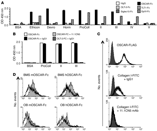
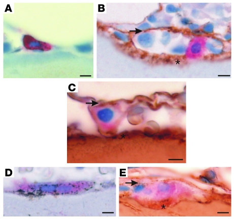
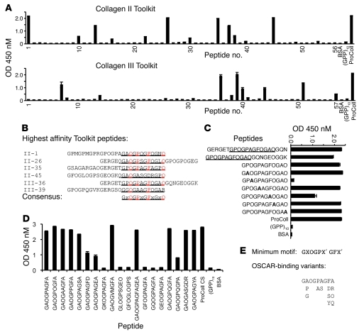
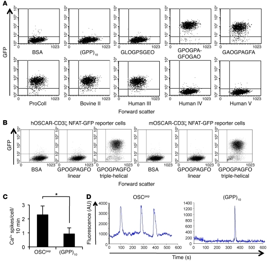
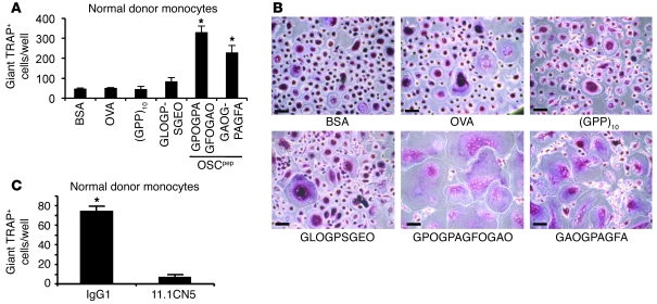
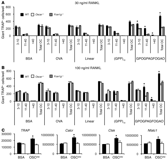
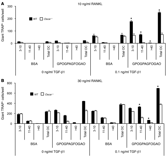
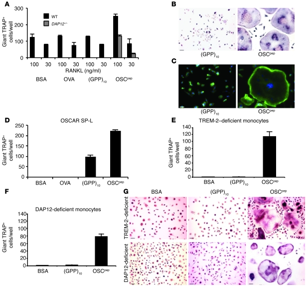
Osteoclasts are terminally differentiated leukocytes that erode the mineralized bone matrix. Osteoclastogenesis requires costimulatory receptor signaling through adaptors containing immunoreceptor tyrosine-based activation motifs (ITAMs), such as Fc receptor common γ (FcRγ) and DNAX-activating protein of 12 kDa. Identification of these ITAM-containing receptors and their ligands remains a high research priority, since the stimuli for osteoclastogenesis are only partly defined. Osteoclast-associated receptor (OSCAR) was proposed to be a potent FcRγ-associated costimulatory receptor expressed by preosteoclasts in vitro, but OSCAR lacks a cognate ligand and its role in vivo has been unclear. Using samples from mice and patients deficient in various ITAM signaling pathways, we show here that OSCAR costimulates one of the major FcRγ-associated pathways required for osteoclastogenesis in vivo. Furthermore, we found that OSCAR binds to specific motifs within fibrillar collagens in the ECM that become revealed on nonquiescent bone surfaces in which osteoclasts undergo maturation and terminal differentiation in vivo. OSCAR promoted osteoclastogenesis in vivo, and OSCAR binding to its collagen motif led to signaling that increased numbers of osteoclasts in culture. Thus, our results suggest that ITAM-containing receptors can respond to exposed ligands in collagen, leading to the functional differentiation of leukocytes, which provides what we believe to be a new concept for ITAM regulation of cytokine receptors in different tissue microenvironments.


Figures

Figure 1
Figure 1. OSCAR is a collagen receptor.
(A) Binding of human Ig-like receptor Fc fusions to BSA or collagens I–V in a solid-phase binding assay (see Methods). A GpVI Fc-fusion protein (GpVI-Fc) was used as positive control for collagen-binding activity, and human IgG (hIgG) was used as negative control. For a description of the other human Ig-like receptor Fc-fusion negative controls, please see Methods and Supplemental Figure 2A. (B) hOSCAR-Fc, preincubated with murine IgG1 or anti-hOSCAR mAb 11.1CN5, binding to BSA or collagen I–III. Solid-phase assay binding data are represented as mean (n = 3) ± SEM. (C) Collagen I–FITC binds to hOSCAR-expressing RBL-2H3 cells (clone 9). RBL-2H3 cells stably expressing hOSCAR-FLAG (white) and untransfected RBL-2H3 cells (dark gray) were stained with FITC-conjugated anti-FLAG mAb and analyzed by flow cytometry. Collagen I–FITC binding to hOSCAR-FLAG transfected RBL-2H3 cells compared with that of untransfected-RBL-2H3 cells. Preincubation with mouse anti-hOSCAR mAb 11.1CN5, but not a mouse IgG1 isotype control mAb, blocks collagen I–FITC binding to hOSCAR-FLAG expressing RBL-2H3 cells. (D) An OSCAR ligand is associated with OBs and stromal cells. Murine OSCAR-Fc (mOSCAR-Fc) and hOSCAR-Fc binding to collagenase-treated (white) or untreated (gray) BMSs or OBs.
Figure 2
Figure 2. Collagens are exposed to preosteoclasts at bone-remodeling sites.
(A) OSCAR (red) is specifically localized in mononuclear cells expressing TRAP (black) on bone surfaces (light blue counterstain). (B) Mononuclear OSCAR+ cells (red) in contact with collagen III (brown) and (C) collagen I (brown) at bone-remodeling sites. (D) Multinucleated TRAP+ cells express OSCAR, which (E) is also located in contact with collagen I at the bone surface. Note the 2 situations in which collagen I and III make contacts with OSCAR+ cells: (a) located below OSCAR+ cells, exposed on bone surfaces (asterisks), and also (b) above OSCAR+ cells, associated with bone-lining cells (arrows) at bone-remodeling sites. The localization of each antigen was validated with 2 independent antibodies. Scale bar: 10 μm.
Figure 3
Figure 3. OSCAR binds to a specific triple-helical motif in collagen.
(A) hOSCAR-Fc binding (y axis, OD 450 nM) to overlapping triple-helical peptides (x axis) from the collagen II and III Toolkits (29, 30). (B) Alignment of triple-helical collagen peptide sequences from Toolkits II and III, displaying highest affinity for hOSCAR-Fc. Predicted hOSCAR collagen-binding consensus is denoted by underlining. Alignment anchor residues are in red. (C) hOSCAR-Fc binding to triple-helical III-36 peptide “halves,” trimmed consensus (underlined), and effect of Alanine scan (bold) through variable X and X′ residues. (D) Effect of various amino acid substitutions through the variable X and X′ residues of the III-36 triple-helical peptide backbone and deletion of the C-terminal triplet on hOSCAR-Fc binding. These data indicate that GPOGPX′GFX, where each proline residue can be substituted by Alanine, is a preferred generic OSCAR-binding motif. Other permissive substitutions remain to be defined. (E) “Minimum” collagen-binding consensus, with alignment of variant hOSCAR-binding residues. Data are represented as mean (n = 3) ± SEM.
Figure 4
Figure 4. The triple-helical conformation of the OSCAR-binding motif in collagen ligands can induce OSCAR signaling.
(A) Dot plots (2,000 events) displaying the response of a hOSCAR-CD3ζ NFAT-GFP reporter cell line to immobilized BSA, (GPP)10, (GPP)5-GLOGPSGEO-(GPP)5, (GPP)5-GPOGPAGFOGAO-(GPP)5, or (GPP)5-GAOGPAGFA-(GPP)5 or collagens I–V (y axis, GFP expression; x axis, forward scatter). (B) Dot plots (10,000 events) displaying the responses (GFP expression) of the hOSCAR-CD3ζ and murine OSCAR-CD3ζ (mOSCAR-CD3ζ) NFAT-GFP reporter cell lines to immobilized BSA; a linear peptide containing the minimal OSCAR-binding sequence GPOGPAGFO (linear); or a triple-helical peptide designed to the minimal OSCAR-binding sequence, (GPP)5-GPOGPAGFO-(GPP)5 (peptide sequences can be found in Supplemental Table 1) (y axis, GFP expression; x axis, forward scatter). (C) Calcium spikes per cell per 10-minute period for human monocytes cultured on either immobilized OSCpep or (GPP)10. Data are represented as mean (n = 5) ± SEM; *P < 0.05. (D) Representative calcium traces for human monocytes cultured on either immobilized OSCpep or (GPP)10.
Figure 5
Figure 5. The OSCAR collagen-binding motif costimulates osteoclastogenesis from human monocytes.
(A) Day 6 RANKL differentiation of human monocytes on the plate-immobilized OSCpep, (GPP)5-GPOGPAGFOGAO-(GPP)5, or (GPP)5-GAOGPAGFA-(GPP)5, or the control protein BSA, or the control peptides OVA or (GPP)10 or (GPP)5-GLOGPSGEO-(GPP)5. Data are represented as mean (n = 3) ± SEM; *P < 0.05 indicates an increase in osteoclasts cultured on OSCpep compared with other culture conditions. (B) TRAP staining of day 6 human osteoclast cultures. Scale bar: 70 μM. (C) Anti-hOSCAR mAb 11.1CN5, but not IgG1, inhibits osteoclastogenesis on immobilized OSCpep, showing the costimulatory action of OSCpep on osteoclastogenesis is hOSCAR specific. Data are represented as mean (n = 3) ± SEM; *P < 0.05.
Figure 6
Figure 6. The OSCAR collagen-binding motif costimulates murine osteoclastogenesis.
(A) RANKL differentiation of wild-type, Oscar–/–, or Fcer1g–/– BMMs cultured on either immobilized OSCpep or control proteins at 30 ng/ml RANKL or (B) 100 ng/ml RANKL. Data are represented as mean (n = 3) ± SEM; *P < 0.05 indicates an increase in osteoclasts cultured on OSCpep compared with all other culture conditions and genotypes. The number of nuclei (e.g., 3–10, 11–40, or >40) per TRAP+ osteoclast (OC), as well as the total number of osteoclasts, was enumerated for each well (x axis). (C) Quantitative RT-PCR expression of osteoclast genes from day 5 osteoclast cultures (black bars, wild type; white bars, Oscar–/–). Data are normalized relative to GAPDH and represented as mean (n = 3) ± SEM; *P < 0.05.
Figure 7
Figure 7. The effects of TGF-β1 and immobilized OSCpep on osteoclastogenesis are additive.
(A) Effect of ±0.1 ng/ml TGF-β1 on the osteoclastogenesis of wild-type or Oscar–/– BMMs cultured on either immobilized BSA or OSCpep at 10 ng/ml RANKL or (B) 30 ng/ml RANKL. The number of nuclei (e.g., 3–10, 11–40, or >40) per TRAP+ osteoclast, as well as the total number of osteoclasts, was enumerated for each well (x axis). Data are represented as mean (n = 3) ± SEM; *P < 0.05 indicates an increase in osteoclasts cultured on OSCpep plus TGF-β1 compared with either BSA with or without TGF-β1 or OSCpep alone.
Figure 8
Figure 8. OSCAR recognition of the collagen motif costimulates osteoclastogenesis of precursors from DAP12–/– mice and TREM-2– and DAP12-deficient patients with NH.
(A) Immobilized OSCpep rescued the osteoclastogenesis of murine DAP12–/– BMMs (day 6) compared with that of wild-type (day 5). (B) TRAP staining (scale bar: 70 μM), (C) DAPI (blue fluorescence), and Phalloidin–Alexa Fluor 488 (green) staining of OSCpep-rescued giant multinucleated DAP12–/– cells (scale bar: 60 μM). (D) Retroviral transduction of mouse OSCAR, long-signal peptide isoform (SP-L), rescued osteoclastogenesis of DAP12–/–Oscar–/– preosteoclasts, showing that the costimulatory signaling and rescue of osteoclastogenesis is OSCAR specific. (E) Effect of immobilized proteins and peptides on osteoclastogenesis of TREM-2–deficient (day 14) or (F) DAP12-deficient monocytes from patients with NH (day 10). (G) TRAP staining of RANKL-differentiated NH monocytes in wells coated with different proteins and peptides (scale bar: 70 μM). Data are represented as mean (n = 3) ± SEM.
Figure 9
Figure 9. OSCAR costimulates a major DAP12-independent pathway for osteoclastogenesis in vivo.
(A) Histology (TRAP, toluidine blue, and von Kossa staining) of the tibia (metaphysis) from DAP12–/–Oscar–/– (DKO) and DAP12–/– mice (scale bar: 100 μM). The bone marrow cavity of DAP12–/–Oscar–/– mice is reduced (toluidine blue staining) and filled with more unresorbed bone (von Kossa) compared with that of DAP12–/– mice. (B) Decrease in TRAP+ osteoclast numbers (osteoclast number/bone surface/mm [N.Oc/BS/mm]), (C) osteoclast size (osteoclast surface/bone surface [Oc.S/BS] [%]), and (D) eroded bone surfaces (eroded surface/bone surface [ES/BS]), with a concomitant increase in (E) trabecular bone volume (bone volume/tissue volume [BV/TV]) in DAP12–/–Oscar–/– mice compared with those in DAP12–/– mice. Data are represented as mean (n = 10) ± SEM; *P < 0.05.

References

    1. Geissmann F, Manz MG, Jung S, Sieweke MH, Merad M, Ley K. Development of monocytes, macrophages, and dendritic cells. Science. 2010;327(5966):656–661. - PMC - PubMed
    1. Gordon S, Martinez FO. Alternative activation of macrophages: mechanism and functions. Immunity. 2010;32(5):593–604. doi: 10.1016/j.immuni.2010.05.007. - DOI - PubMed
    1. Martin TJ. Paracrine regulation of osteoclast formation and activity: milestones in discovery. J Musculoskelet Neuronal Interact. 2004;4(3):243–253. - PubMed
    1. Kong YY, et al. Activated T cells regulate bone loss and joint destruction in adjuvant arthritis through osteoprotegerin ligand. Nature. 1999;402(6759):304–309. - PubMed
    1. Paloneva J, et al. Loss-of-function mutations in TYROBP (DAP12) result in a presenile dementia with bone cysts. Nat Genet. 2000;25(3):357–361. doi: 10.1038/77153. - DOI - PubMed

Publication types

MeSH terms