. 2011 Sep;121(9):3623-34.
doi: 10.1172/JCI46323. Epub 2011 Aug 15.

Human Merkel cell polyomavirus small T antigen is an oncoprotein targeting the 4E-BP1 translation regulator

Affiliations

Human Merkel cell polyomavirus small T antigen is an oncoprotein targeting the 4E-BP1 translation regulator

Masahiro Shuda et al. J Clin Invest. 2011 Sep.

Abstract

Merkel cell polyomavirus (MCV) is the recently discovered cause of most Merkel cell carcinomas (MCCs), an aggressive form of nonmelanoma skin cancer. Although MCV is known to integrate into the tumor cell genome and to undergo mutation, the molecular mechanisms used by this virus to cause cancer are unknown. Here, we show that MCV small T (sT) antigen is expressed in most MCC tumors, where it is required for tumor cell growth. Unlike the closely related SV40 sT, MCV sT transformed rodent fibroblasts to anchorage- and contact-independent growth and promoted serum-free proliferation of human cells. These effects did not involve protein phosphatase 2A (PP2A) inhibition. MCV sT was found to act downstream in the mammalian target of rapamycin (mTOR) signaling pathway to preserve eukaryotic translation initiation factor 4E-binding protein 1 (4E-BP1) hyperphosphorylation, resulting in dysregulated cap-dependent translation. MCV sT-associated 4E-BP1 serine 65 hyperphosphorylation was resistant to mTOR complex (mTORC1) and mTORC2 inhibitors. Steady-state phosphorylation of other downstream Akt-mTOR targets, including S6K and 4E-BP2, was also increased by MCV sT. Expression of a constitutively active 4E-BP1 that could not be phosphorylated antagonized the cell transformation activity of MCV sT. Taken together, these experiments showed that 4E-BP1 inhibition is required for MCV transformation. Thus, MCV sT is an oncoprotein, and its effects on dysregulated cap-dependent translation have clinical implications for the prevention, diagnosis, and treatment of MCV-related cancers.


Figures
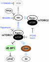
Figure 1
Figure 1. Akt-mTOR pathway.
Activities of Akt, mTORC1 (Raptor complex), mTORC2 (Rictor complex), and S6K kinases are shown. mTORC1 phosphorylates and inhibits 4E-BP1, which prevents 4E-BP1 from sequestering the eIF4E cap-dependent translation factor. Kinase inhibitors used in this study are shown in blue, and the respective targets are designated by dotted lines.
Figure 2
Figure 2. MCV sT antigen protein is commonly expressed in MCC.
Immunohistochemical staining of adjacent slides from 2 MCC cases with mouse mAbs to MCV sT (CM5E1 antibody) and MCV LT (CM2B4 antibody) is shown. MCV T antigens were localized only to tumor cells and were not detected in nontumor interstitial tissues. (A and B) MCC case MCCR10-123, in which both sT and LT were expressed. (D and E) MCC case MCC344, with abundant sT protein (D) but no LT protein (E) expression. (C and F) SV40 LT antigen (PAb419 antibody) was used as a negative control and was not positive for either MCC case. Original magnification, ×100; ×1,000 (insets). (G) Blinded analysis of 51 consecutive CK20-positive MCC tumors stained for sT and LT. MCV sT positivity in MCCs, when present, ranged in intensity from slight to robust.
Figure 3
Figure 3. MCV sT expression is required for growth, but not survival, of MCV-positive MCC cells.
(A) Knockdown with pan-T1 shRNA in MCV-positive MKL-1 cells reduced both LT and sT antigen expression, whereas knockdown targeting with sT1 reduced sT expression alone. sh ctrl, control shRNA. (B) Cell proliferation was reduced in MKL-1 cells, but not MCV-negative UISO cells, with both pan-T1 and sT1 shRNA lentivirus transduction (normalized by mean OD values on day 1). (C) S and G2/M phase cell cycle entry, measured by BrdU uptake (red), was markedly reduced by pan-T1 lentiviral knockdown compared with control shRNA for MKL-1 cells, but not UISO cells. Reproducible but modest cell cycle inhibition occurred with sT knockdown in MCV-positive MCC cell lines. Percent cells with BrdU incorporation is indicated within the histograms. PI, propidium iodide. (D) sT knockdown did not cause MCC cell death. LDH release was elevated for MKL-1 cells transduced with pan-T1 shRNA lentivirus, indicating increased cell death. No increase in LDH release occurred after sT1 shRNA transduction. Average values for 3 independent experiments are shown.
Figure 4
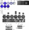
Figure 4. MCV sT induces PP2A-binding and DnaJ domain–independent transformation of Rat-1 cells.
(A) Lentiviral expression of MCV sT generated dense foci formation in Rat-1 cells compared with empty vector or full-length MCV LT cDNA. Tumor-derived LT antigens (LT.339 and LT.350) also did not increase focus formation. Mutations in the sT PP2A-binding (sT.R7A and sT.L142A) or DnaJ (sT.D44N) domains did not affect focus formation. Number of foci per dish is indicated. (B) Phase-contrast images of foci for empty vector– and MCV sT–expressing Rat-1 cells. Original magnification, ×40. (C) MCV sT, but not LT, induced anchorage-independent growth of Rat-1 cells in soft agar; this was unaffected by PP2A-binding (sT.R7A and sT.L142A) or DnaJ (sT.D44N) domain mutations. Colonies observed in 6-well triplicates were counted to determine average ± SD colonies per well. Colonies in wells (dark spots) and photomicrographs of typical fields are shown for each condition. Original magnification, ×200. (D) LT and sT expression in Rat-1 cells from C were detected by immunoblotting with CM2B4 and CM5E1, respectively. (E) MCV sT interacted with the Aα subunit of PP2A. HA-tagged PP2A Aα subunit was overexpressed in 293 cells, together with wild-type sT cDNA or with PP2A- or DnaJ-binding mutant cDNAs, and immunoprecipitated with HA antibody. Pulldown of sT protein was detected using anti-sT antibody (CM5E1).
Figure 5
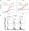
Figure 5. MCV sT promotes serum-independent human BJ-TERT cell proliferation.
(A) sT protein was transduced by lentiviral vector into immortalized BJ-TERT cells, and cell proliferation was determined in 10% FCS or no serum using a Wst-1 colorimetric cell proliferation assay. sT accelerated BJ-TERT cell growth in the presence of serum and prevented proliferation arrest in the absence of serum (normalized by mean OD values on day 1 for 2 independent experiments performed in triplicate). Mutation of the sT binding site to PP2A (sT.L142A) retained cell proliferation in both the presence and the absence of serum. (B) Cell cycle profiles for BJ-TERT cells expressing sT antigens in 0% and 10% FCS. In the absence of serum, greater than 95% of BJ-TERT cells transduced with the empty vector arrested in G1, whereas substantial fractions of cells expressing wild-type sT (7%) or sT.L142A (11%) continued to transit through the cell cycle.
Figure 6
Figure 6. MCV sT promotes 4E-BP1 S65 hyperphosphorylation and cap-dependent translation.
(A) MCV sT promoted δ 4E-BP1 hyperphosphorylation at S65; this phosphorylation was mediated by mTORC1 and could be inhibited by long-term raptor knockdown. 293 cells were stably transduced with Raptor shRNA lentivirus (shRap) and transfected with sT or empty vector on day 5 after transduction. (B) MCV sT prevented loss of hyperphosphorylated 4E-BP1 S65 during short-term mTORC1 inhibition with rapamycin. 293 cells transfected with empty vector or MCV sT were treated with 50 nM rapamycin for up to 1 hour. Basal phosphorylation at T37 and T46 was unaffected by rapamycin treatment or sT expression. (C) MCV sT prevented loss of 4E-BP1 during short-term mTORC1 and mTORC2 inhibition with Torin1 and PP242. 293 cells, with or without sT expression, were treated with Torin1 (500 nM) or PP242 (5 μM) for 6 hours. 4E-BP1 was almost completely dephosphorylated after drug treatment in the absence of sT expression. When sT was expressed, the δ S65 form was preserved. (D) Both wild-type sT and PP2A-binding sT mutants promoted rapamycin-resistant 4E-BP1 phosphorylation in 293 cells treated with 50 nM rapamycin for 1 hour.
Figure 7
Figure 7. MCV sT knockdown in MCV-infected MCC cells reduces 4E-BP1 hyperphosphorylation and inactivates cap-dependent translation initiation complex formation.
(A) sT and pan-T antigen knockdown reduced 4E-BP1 γ S65 hyperphosphorylation in MCV-positive MKL-1 cells, but not MCV-negative UISO cells. Priming site T37/T46 phosphorylation was preserved after sT antigen knockdown, whereas secondary sites of phosphorylation (S65 and T70) were markedly reduced. (B) Knockdown of either pan-T antigen or sT (by pan-T1 or sT1, respectively) in MKL-1 cells inhibited eIF4G binding to 7mGTP sepharose beads in parallel with loss of 4E-BP1 S65 phosphorylation. Control shRNA–infected MKL-1 cells treated with PP242 are shown as a positive control. Despite partial preservation of 4E-BP1 phospho-S65 (arrowhead) in MKL-1 cells, eIF4E and eIF4G binding was reduced after PP242 treatment. Lanes were run on the same gel but were noncontiguous (white lines).
Figure 8
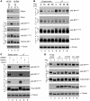
Figure 8. sT promotes steady-state phosphorylation of other mTORC1 downstream molecules.
(A) sT and PP2A-binding mutants of MCV sT (sT.R7A and sT.L142A) increased steady-state phosphorylation of the S6K kinase at amino acid T389 and T421/S424, its S6 ribosomal protein substrate at S235/S236, and the 4E-BP2 protein (arrowheads). Phosphorylation promoted by sT was relatively resistant to 1 hour rapamycin treatment, except at the S6K T389 site. α-Tubulin was used as a loading control. (B) sT, sT.R7A, and sT.L142A did not promote S473 Akt phosphorylation. sT did not prevent PP242 or Torin1 from blocking mTOR-dependent Akt phosphorylation. (C) PP2A targeting by MCV sT did not promote Akt phosphorylation. (D) MKL-1 cells were transduced with sT1, pan-T, or scrambled shRNA vector lentiviruses for 6 days, and then cell lysates were harvested for immunoblotting. The PI3K inhibitor LY294002 and the Akt inhibitor MK2206 were used as positive controls to show constitutive Akt activity in MKL-1 cells. Akt phosphorylation was increased by both pan-T1 shRNA and sT1 shRNA knockdown.
Figure 9
Figure 9. Role of 4E-BP1 phosphorylation in sT-induced transformation.
(A) Rat-1 cells were stably transduced with empty vector, wild-type 4E-BP1, or constitutively active 4E-BP1AA. Empty vector or MCV sT was then cotransduced into the cells without selection, and they were grown in soft agar for 3 weeks. Colonies were stained with crystal violet, and the colony number per high-power field was determined. (B) Phase-contrast images of sT-induced Rat-1 cell colonies in soft agar. Original magnification, ×40.
Figure 10
Figure 10. Proposed mechanism of action for MCV sT in cell transformation.
MCV sT preserves 4E-BP1 hyperphosphorylation, most likely by preventing hyperphosphorylated 4E-BP1 turnover, which increases cap-dependent protein translation in Merkel cell cancers. The MCV protein does not markedly affect priming 4E-BP1 phosphorylation at residues T37 and T46 and does not directly induce 4E-BP1 phosphorylation.

Similar articles

Cited by

References

    1. Gross L. A filterable agent, recovered from Ak leukemic extracts, causing salivary gland carcinomas in C3H mice. Proc Soc Exp Biol Med. 1953;83(2):414–421. - PubMed
    1. Lane DP, Crawford LV. T antigen is bound to a host protein in SV40-transformed cells. Nature. 1979;278(5701):261–263. doi: 10.1038/278261a0. - DOI - PubMed
    1. Linzer DI, Levine AJ. Characterization of a 54K dalton cellular SV40 tumor antigen present in SV40-transformed cells and uninfected embryonal carcinoma cells. Cell. 1979;17(1):43–52. doi: 10.1016/0092-8674(79)90293-9. - DOI - PubMed
    1. DeCaprio JA, et al. SV40 large tumor antigen forms a specific complex with the product of the retinoblastoma susceptibility gene. Cell. 1988;54(2):275–283. doi: 10.1016/0092-8674(88)90559-4. - DOI - PubMed
    1. Eckhart W, Hutchinson MA, Hunter T. An activity phosphorylating tyrosine in polyoma T antigen immunoprecipitates. Cell. 1979;18(4):925–933. doi: 10.1016/0092-8674(79)90205-8. - DOI - PubMed

Publication types

MeSH terms

Substances