. 2011 Sep;121(9):3442-55.
doi: 10.1172/JCI45390. Epub 2011 Aug 15.

miR-107 promotes tumor progression by targeting the let-7 microRNA in mice and humans

Affiliations

miR-107 promotes tumor progression by targeting the let-7 microRNA in mice and humans

Pai-Sheng Chen et al. J Clin Invest. 2011 Sep.

Erratum in

Abstract

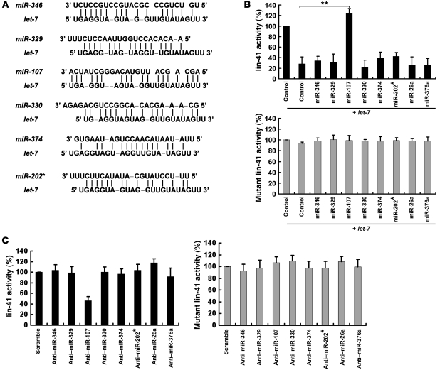
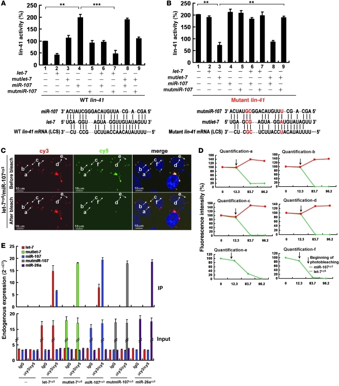
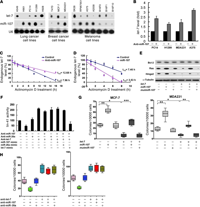
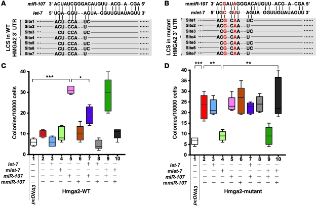
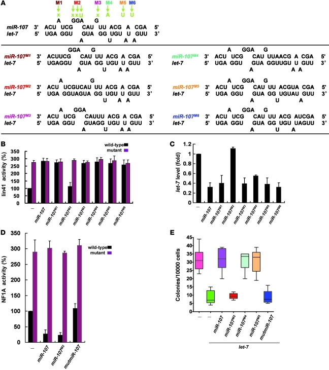
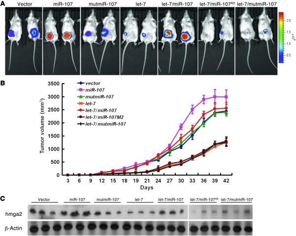
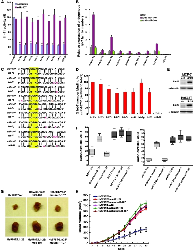
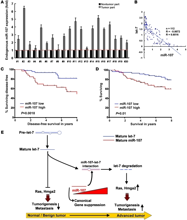
MicroRNAs (miRNAs) influence many biological processes, including cancer. They do so by posttranscriptionally repressing target mRNAs to which they have sequence complementarity. Although it has been postulated that miRNAs can regulate other miRNAs, this has never been shown experimentally to our knowledge. Here, we demonstrate that miR-107 negatively regulates the tumor suppressor miRNA let-7 via a direct interaction. miR-107 was found to be highly expressed in malignant tissue from patients with advanced breast cancer, and its expression was inversely correlated with let-7 expression in tumors and in cancer cell lines. Ectopic expression of miR-107 in human cancer cell lines led to destabilization of mature let-7, increased expression of let-7 targets, and increased malignant phenotypes. In contrast, depletion of endogenous miR-107 dramatically increased the stability of mature let-7 and led to downregulation of let-7 targets. Accordingly, miR-107 expression increased the tumorigenic and metastatic potential of a human breast cancer cell line in mice via inhibition of let-7 and upregulation of let-7 targets. By mutating individual sites within miR-107 and let-7, we found that miR-107 directly interacts with let-7 and that the internal loop of the let-7/miR-107 duplex is critical for repression of let-7 expression. Altogether, we have identified an oncogenic role for miR-107 and provide evidence of a transregulational interaction among miRNAs in human cancer development.


Figures

Figure 1
Figure 1. miR-107 suppresses let-7 function.
(A) Potential duplexes formed between let-7a and candidate miRNAs. (B) A549 cells were cotransfected with lin-41 and the indicated antagomirs. Firefly luciferase reporter activity was normalized to renilla luciferase. MiR-26a and miR-376a were included as nonspecific miRNAs. Control cells in lane 1 carried pcDNA6.2-EmGFP empty vectors and were transfected with the lin-41 reporter. (C) Depletion of endogenous miR-107 results in enhanced let-7–suppressed lin-41 expression. MDA231 cells were cotransfected with lin-41 and the miRNA antagomirs indicated. Firefly luciferase reporter activity was normalized to renilla luciferase. MiR-26a and miR-376a were included as nonspecific miRNAs. Scramble oligo-transfected cells were used as the control group. *P < 0.05; **P < 0.01. Data are presented as mean ± SD.
Figure 2
Figure 2. Direct interaction between miR-107 and let-7.
(A) Design of constructs containing wild-type (A) and mutant (B) miR-107, let-7, and lin-41. The 3′ UTR of lin-41 was appended to the luciferase open reading frame (Luc). The let-7 complementary sites are indicated. miR-107, let-7, lin-41, and their mutants were transfected into H1299 cells and assayed for luciferase activity. **P < 0.01; ***P < 0.001. (C) In vivo cellular localization of miR-107 and let-7a in A549 cells after photobleaching. Cells with colocalization of miR-107/let-7a observed by confocal microscopy (Supplemental Figure 2B) were bleached repeatedly by λ= 633 nm, and the fluorescence intensity of cy3-labeled miR-107 was subsequently monitored at λ= 575–615 nm before and after photobleaching. (D) The FRET efficiency in each location shown in C. a–d: colocalized complexes; e, f: complexes without colocalization. (E) The interaction between miR-107 and let-7a as assessed by immunoprecipitation. A549 cells expressing cy3–miR-107 and cy5–let-7a were subjected to immunoprecipitation using anti-cy3/cy5 antibodies and then assayed by qRT-PCR. Data are presented as mean ± SD.
Figure 3
Figure 3. miR-107 promotes let-7 degradation and antagonizes let-7–suppressed AIG in human cancer cell lines.
(A) Expression of miR-107 and let-7 in human cancer cell lines. Total RNA (20 μg) was isolated and assayed by Northern blotting with specific probes for let-7a and miR-107. (B) Effect of miR-107 on let-7a expression. Total RNA was isolated from cells transfected with miR-107 antagomir and then assayed by qRT-PCR. U6 was used as an internal control, and the fold change of the let-7a level was normalized with the paired untreated group. (C) An miR-107 antagomir was introduced into H1299 cells in the presence of 1 μg/ml of actinomycin D. Total RNA was isolated and assayed by qRT-PCR for mature let-7a. (D) miR-107 was introduced into A549 cells in the presence of 1 μg/ml of actinomycin D. Total RNA was isolated and mature let-7a was assayed for by qRT-PCR. (E) Forced expression of miR-107 induced the expression of both Ras and HMGA2. H1299 cells were transfected with indicated miRNAs, and Ras and HMGA2 levels were assayed by Western blotting. (F) Dual-luciferase assays were performed to determine the lin-41 expression in the presence of miR-107, let-7a mimics, or antagomirs. (G and H) MiRNA mimics or antagomirs were introduced into MCF-7 or MDA231 cells and effects of miR-107 on colony formation were determined. The indicated miRNAs or antagomirs were transfected into MCF-7 (G, H), MDA-MB-231 (G), and H1299 (H) cells. Soft agar assays were performed over 14 days. *P < 0.05; **P < 0.01; ***P < 0.001. Data are presented as mean ± SD.
Figure 4
Figure 4. miR-107–mediated induction of the let-7–suppressed tumorigenicity requires direct base-pairing of miR-10:let-7:HMGA2.
(A and B) HMGA2 constructs used in the AIG assays. The let-7 complementary and mutated sites are indicated. The mutated HMGA2 3′ UTR site had 2 point substitutions that disrupted pairing to let-7 but allowed pairing to mlet-7. LCS, let-7 complementary site. (C and D) The requirement of pairing between miR-107 and let-7 on colony formation. The indicated miRNA-expressing constructs were transfected into wild-type or mutated HMGA2-expressing MCF-7 cells. Cells were then assayed for colony formation as described above. *P < 0.05; **P < 0.01; ***P < 0.001. Data are presented as mean ± SD.
Figure 5
Figure 5. The internal loop of the miR-107–let-7 duplex is crucial for their activities.
(A) Duplex structures formed between mutant forms of miR-107 and let-7a (bottom). Design of mutated miR-107 constructs. The top panel shows the wild-type miR-107–let-7a duplex structure, which exhibits several mismatched regions; mutations in miR-107 eliminating those mismatches are indicated (M1–M6). (B and C) The internal loop is required for miR-107 to antagonize let-7 function. The effects of structure-based miR-107 mutants on lin-41 activity (B) and let-7a levels (C) were measured in T47D cells. Luciferase activity of wild-type lin-41 in the control group served as the reference (100%). (D) Effect of the mutant forms of miR-107 on NF1A suppression. The activity of NF1A, a known mRNA target of miR-107, was measured using a luciferase reporter assay. The wild type and mutant refer to the NF1A constructs used in the reporter assay. (E) Requirement of miR-107’s internal loop for in vitro tumorigenesis. The effect of miR-107M2 mutants on colony formation, as measured by AIG assays. Data are presented as mean ± SD.
Figure 6
Figure 6. Effects of miR-107, let-7, and their mutants on tumorigenesis and metastasis in an animal model.
(A and B) The effects of expression of miR-107, let-7a, and their mutants on orthotropic tumor growth were assessed by in vivo image detection. (A) The tumor burden was measured and calculated directly every 3 days, and the results were plotted as tumor volume over time (B). (C) Expression of HMGA2 in orthotropic tumor tissues was measured by Western blotting. Blot lanes were run on the same gel but were noncontiguous (white line). Data are presented as mean ± SD.
Figure 7
Figure 7. Regulation of let-7 family members by miR-107.
(A) Effect of miR-107 on lin-41 expression suppressed by different let-7 members. H661 cells were cotransfected with lin-41 and miR-107 in the presence or absence of let-7 member overexpression. Firefly luciferase reporter activity was normalized to renilla luciferase. (B) Expression of let-7 family members in miR-107 knockdown cells. The indicated antagomirs were transfected into MCF-7 cells and then assayed by real-time RT-PCR. (C) Duplex between miR-107 and let-7 family members. The yellow box represents the conserved sequence within the regions of internal loop. Red, residues differ from let-7a. (D) Binding between miR-107 and let-7 family members. miR-107cy3 was transfected into MCF-7 cells. The binding between miR-107cy3 and endogenous let-7 family members was determined by real-time RT-PCR. Percentage of let-7 members binding to miR-107cy3 was normalized with control. (E) Successful overexpression of Lin28 in MCF-7 and Hs578T cells. MCF-7 and Hs578T cells were stably transfected with pBabe-hLin28B. Lysates were collected and assayed by Western blot. (F) Effect of miR-107 on anchorage-independent growth in Lin28-overexpressed (let-7 knockdown) cells. MCF-7 and Hs578T cells were transfected with indicated miRNAs in the presence or absence of Lin28 overexpression. Soft agar assays were performed for 14 days. (G and H) Effect of miR-107 on in vivo tumor growth in Lin28-overexpressed cells. Hs578T/Vec and Hs578T/Lin28 cells were transfected with indicated miRNAs in the presence or absence of Lin28 overexpression. The tumor burden was measured and calculated every 3 days, and the results were plotted as tumor volume over time. Data are presented as mean ± SD.
Figure 8
Figure 8. miR-107 is overexpressed in human breast cancer tissues and is related to poor prognosis.
(A) Expression of miR-107 in normal/tumor portions of breast cancer tissues. We isolated RNAs from paired tumor and nontumor samples. Expression of miR-107 was detected by qRT-PCR. The level of miR-107 in nontumor tissue was used as a reference (= 1), and the relative fold expression in the tumor tissue was determined. (B) The correlation between miR-107 and let-7a levels in human breast cancer tissues. qRT-PCR was used to detect expression of miR-107 and let-7a. Each point on the graph corresponds to the relative expression levels from an individual patient. We normalized let-7a and miR-107 levels with U6 to calculate their relative expression levels. We used the averaged expression level of all patients as cutoff value. (C and D) miR-107 level predicts poor clinical outcome in patients with breast cancer. The log-rank test (2-sided) was used to compare differences between groups. The Kaplan-Meier curves show analyses of disease-free (C) and overall (D) survival in patients with breast cancer. (E) Working model of the miR-107–let-7 interaction during cancer progression. Data are presented as mean ± SD.

References

    1. Bartel DP. MiRNAs: genomics, biogenesis, mechanism, and function. Cell. 2004;116(2):281–297. doi: 10.1016/S0092-8674(04)00045-5. - DOI - PubMed
    1. Mattick JS, Makunin IV. Non-coding RNA. Hum Mol Genet. 2006;15 spec no 1:R17–R29. - PubMed
    1. Johnson SM, et al. RAS is regulated by the let-7 miRNA family. Cell. 2005;120(5):635–647. doi: 10.1016/j.cell.2005.01.014. - DOI - PubMed
    1. Takamizawa J, et al. Reduced expression of the let-7 miRNAs in human lung cancers in association with shortened postoperative survival. Cancer Res. 2004;64(11):3753–3756. doi: 10.1158/0008-5472.CAN-04-0637. - DOI - PubMed
    1. Yu F, et al. let-7 regulates self renewal and tumorigenicity of breast cancer cells. Cell. 2007;131(6):1109–1123. - PubMed

Publication types