. 2011 Aug 14;18(9):971-6.
doi: 10.1038/nsmb.2099.

The pseudokinase domain of JAK2 is a dual-specificity protein kinase that negatively regulates cytokine signaling

Affiliations

The pseudokinase domain of JAK2 is a dual-specificity protein kinase that negatively regulates cytokine signaling

Daniela Ungureanu et al. Nat Struct Mol Biol. .

Abstract

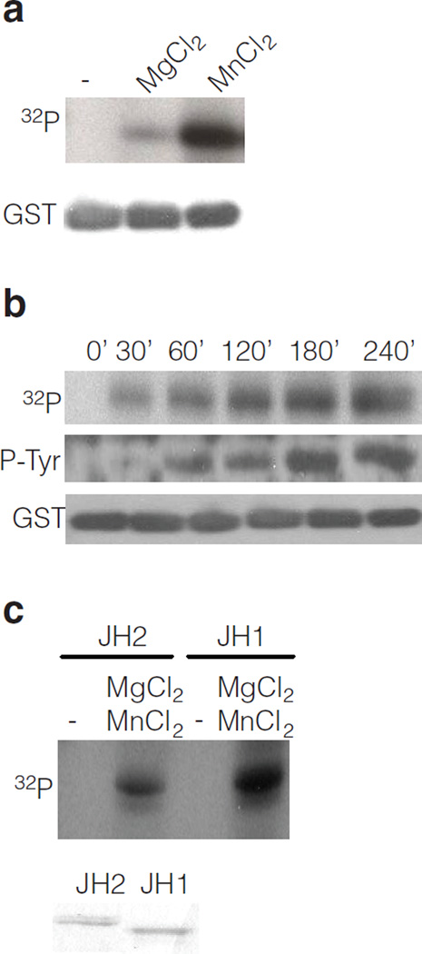
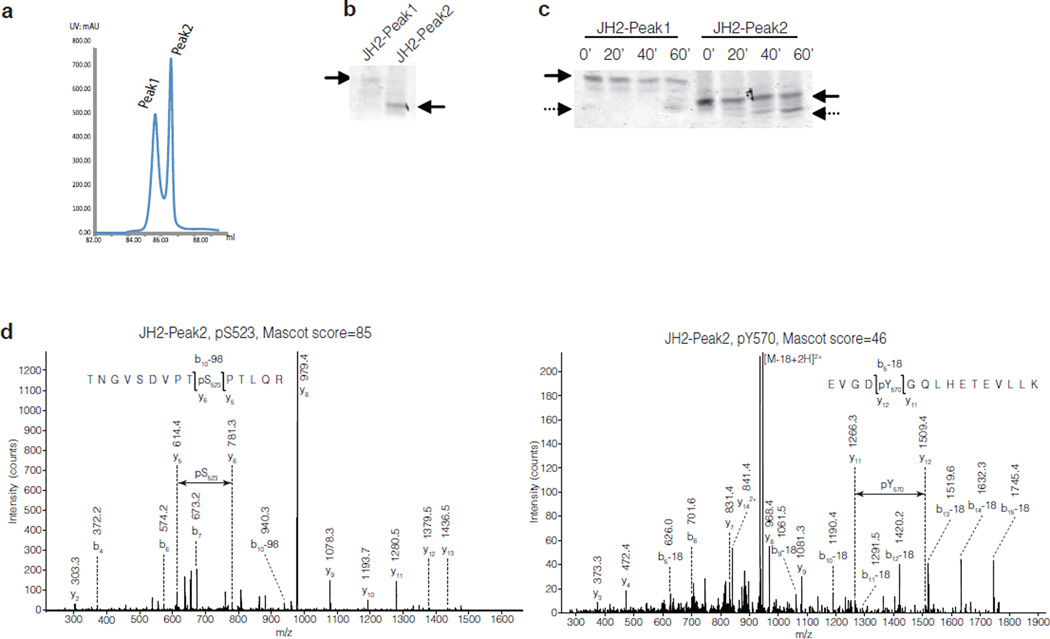
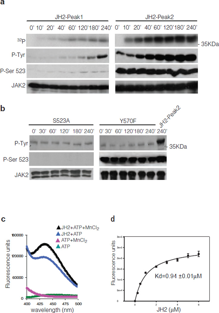
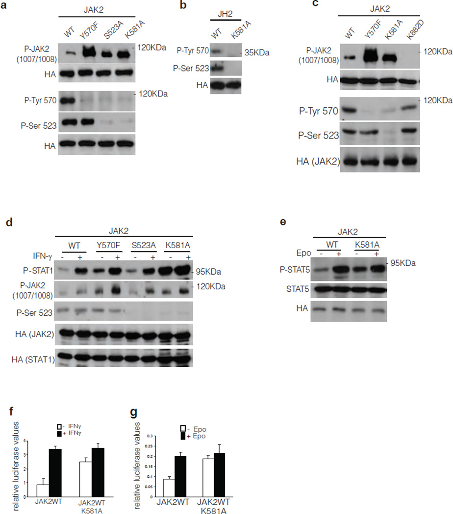
Human JAK2 tyrosine kinase mediates signaling through numerous cytokine receptors. The JAK2 JH2 domain functions as a negative regulator and is presumed to be a catalytically inactive pseudokinase, but the mechanism(s) for its inhibition of JAK2 remains unknown. Mutations in JH2 lead to increased JAK2 activity, contributing to myeloproliferative neoplasms (MPNs). Here we show that JH2 is a dual-specificity protein kinase that phosphorylates two negative regulatory sites in JAK2: Ser523 and Tyr570. Inactivation of JH2 catalytic activity increased JAK2 basal activity and downstream signaling. Notably, different MPN mutations abrogated JH2 activity in cells, and in MPN (V617F) patient cells phosphorylation of Tyr570 was reduced, suggesting that loss of JH2 activity contributes to the pathogenesis of MPNs. These results identify the catalytic activity of JH2 as a previously unrecognized mechanism to control basal activity and signaling of JAK2.


Figures

Figure 1
Figure 1
Identification of JAK2 JH2 catalytic activity in vitro. (a) In vitro kinase assay with purified JAK2 GST-JH2 with [32P] γ-ATP in the absence or presence of divalent cations. (b) Time-course kinase assay with purified JAK2 GST-JH2 in the presence of [γ- 32P] ATP or unlabeled ATP. (c) Autoradiography of kinase assay (30 min) using purified JAK2 JH2 and JH1 domain and [γ-32P] ATP, in the absence or presence of cations. Coomassie staining shows the protein levels of JH1 and JH2.
Figure 2
Figure 2
Identification of phosphorylated residues in JAK2 JH2. (a) Chromatogram of JAK2 JH2 purification showing the peaks from anion-exchange chromatography. (b) Coomassie staining of a native-gel electrophoresis of JH2-Peak1 and JH2-Peak2 proteins. (c) Coomassie staining of a native-gel electrophoresis of purified JH2-Peak1 and JH2- Peak2 after kinase reaction. (d) MS-MS spectra of the phosphorylated residues in JAK2 JH2-Peak2 4h kinase assay. Left: JH2-Peak2 is stoichiometrically phosphorylated at Ser523. Right: JH2-Peak2 is partially phosphorylated at Tyr570.
Figure 3
Figure 3
Analysis of JAK2 JH2 autophosphorylation and ATP binding activity. (a) Timecourse kinase assay of purified JH2-Peak1 and JH2-Peak2. (b) Time-course kinase assay of purified JH2 S523A and Y570F mutants compared to JH2-Peak2. (c) Fluorescence measurement of ATP binding assay of JAK2 JH2-Peak2. (d) Kd measurement of mant-ATP binding to JH2-Peak2. Graph, mean ± s.d. of three independent experiments.
Figure 4
Figure 4
Analysis of JAK2 signaling in mammalian cells. (a, c) Phosphorylation of JAK2WT and mutants thereof in JAK2-deficient γ2A cells. HA-tagged JAK2 proteins were immunoprecipitated with anti-HA antibody and JAK2 phosphorylation is shown by Western blotting. Anti-HA Western blots show protein levels for each independent experiment. (b) Phosphorylation of JAK2 JH2 in γ2A cells. (d, e) Phosphorylation of STAT1 in response to IFN-γ stimulation and phosphorylation of STAT5 in response to Epo stimulation in γ2A cells. (f) Effect of JAK2 K581A mutation on STAT1 transcription activation using IF–γ-dependent GAS luciferase reporter. Graph, mean ± s.d. of three independent experiments, P < 0.05. (g) Effect of JAK2 K581A mutation on STAT5 transcription activation using SPI-Luc2 luciferase reporter. The basal JAK2 WT activity was set to 1 for all experiments, graph, mean ± s.d. of six independent experiments are shown, P < 0.05.
Figure 5
Figure 5
Phosphorylation of different JAK2 MPN mutants. (a) Phosphorylation of JAK2WT and MPN mutants in JAK2-deficient γ2A cells. (b) Phosphorylation of JAK2 JH2 in γ2A cells.

Comment in

  • JH2 is active!
    Baumann K. Baumann K. Nat Rev Mol Cell Biol. 2011 Aug 23;12(9):550. doi: 10.1038/nrm3183. Nat Rev Mol Cell Biol. 2011. PMID: 21860390 No abstract available.

References

    1. Gadina M, et al. Signaling by type I and II cytokine receptors: ten years after. Curr. Opin. Immunol. 2001;13:363–373. - PubMed
    1. Imada K, Leonard WJ. The Jak-STAT pathway. Mol. Immunol. 2000;37:1–11. - PubMed
    1. Silvennoinen O, et al. Structure of the murine Jak2 protein-tyrosine kinase and its role in interleukin 3 signal transduction. Proc. Natl. Acad. Sci. U. S. A. 1993;90:8429–8433. - PMC - PubMed
    1. Yamaoka K, et al. The Janus kinases (Jaks) Genome Biol. 2004;5:253. - PMC - PubMed
    1. Ishida-Takahashi R, et al. Phosphorylation of Jak2 on SeR523) inhibits Jak2-dependent leptin receptor signaling. Mol. Cell. Biol. 2006;26:4063–4073. - PMC - PubMed

Methods references

    1. Shevchenko A, Tomas H, Havlis J, Olsen JV, Mann M. In-gel digestion for mass spectrometric characterization of proteins and proteomes. Nat. Protoc. 2006;1:2856–2860. - PubMed
    1. Thingholm TE, Jorgensen TJ, Jensen ON, Larsen MR. Highly selective enrichment of phosphorylated peptides using titanium dioxide. Nat. Protoc. 2006;1:1929–1935. - PubMed
    1. Ye J, et al. Optimized IMAC-IMAC Protocol for Phosphopeptide Recovery from Complex Biological Samples. J. Proteome Res. 2010;9:3561–3573. - PubMed

Publication types